Hyperargininemia
Apr. 14, 2026
MedLink, LLC
3525 Del Mar Heights Rd, Ste 304
San Diego, CA 92130-2122
Toll Free (U.S. + Canada): 800-452-2400
US Number: +1-619-640-4660
Support: service@medlink.com
Editor: editor@medlink.com
ISSN: 2831-9125
Toll Free (U.S. + Canada): 800-452-2400
US Number: +1-619-640-4660
Support: service@medlink.com
Editor: editor@medlink.com
ISSN: 2831-9125
Nearly 3,000 illustrations, including video clips of neurologic disorders.
Every article is reviewed by our esteemed Editorial Board for accuracy and currency.
Full spectrum of neurology in 1,200 comprehensive articles.
Listen to MedLink on the go with Audio versions of each article.
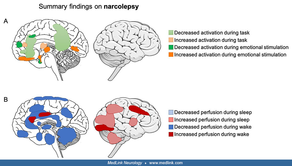
(A) Summary of findings from fMRI studies in narcolepsy. During winning on a gambling task, activity is reduced in the bilateral nucleus accumbens and the ventromedial prefrontal cortex, and activity is increased in the putamen and inferior lateral frontal cortex. During an encoding and recognition task, there is more deactivation within the default mode network. In response to the presentation of humorous images, decreased activation is present in the right hypothalamus, medial prefrontal cingulate cortex, nucleus accumbens, and hypothalamus. Increased activity is present in the right inferior frontal gyri and amygdala. (B) Summary of findings from PET and SPECT studies in narcolepsy. During sleep, activity in temporoparietal regions of the cortex and cerebellum is increased; perfusion within parietal regions is increased during REM sleep. During wakefulness, there is decreased perfusion in the brainstem, cerebellum, caudate nucleus, subcallosal gyrus, and cingulate gyrus extending along the corpus callosum. Glucose metabolism is decreased in the parahippocampal gyrus, bilateral paracentral areas, bilateral hypothalami, thalamic nuclei, and frontal-parietal cortices. Hypermetabolism is present in the anterior and midcingulate cortex, the right cuneus, and the lingual gyrus. (Contributed by Dr. Nathan Cross and Dr. Florence Pomares.)