Stroke & Vascular Disorders
Amaurosis fugax
Jan. 20, 2026
MedLink, LLC
3525 Del Mar Heights Rd, Ste 304
San Diego, CA 92130-2122
Toll Free (U.S. + Canada): 800-452-2400
US Number: +1-619-640-4660
Support: service@medlink.com
Editor: editor@medlink.com
ISSN: 2831-9125
Toll Free (U.S. + Canada): 800-452-2400
US Number: +1-619-640-4660
Support: service@medlink.com
Editor: editor@medlink.com
ISSN: 2831-9125
Nearly 3,000 illustrations, including video clips of neurologic disorders.
Every article is reviewed by our esteemed Editorial Board for accuracy and currency.
Full spectrum of neurology in 1,200 comprehensive articles.
Listen to MedLink on the go with Audio versions of each article.
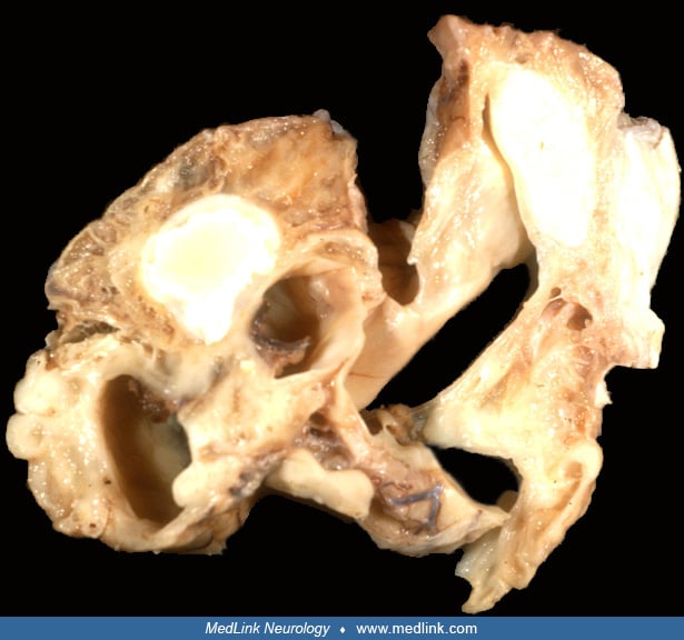
The patient was a 5-and-a-half-year-old male with history of HSV-2 encephalitis at 10 months. The brain was reduced in volume, with extensive involvement of the temporal and frontal lobes. Coronal section shows extensive multiloculated cavitation within the frontal and temporal lobes. Clinical details of the case have been published in: Feldman RA, Shende M. Herpes simplex virus encephalitis simulating a frontoparietal convexity neoplasm. Surg Neurol 1975;3:329-32. (Contributed by R Kim.)