


Developmental Malformations
Waardenburg syndrome
Jul. 18, 2025
MedLink, LLC
3525 Del Mar Heights Rd, Ste 304
San Diego, CA 92130-2122
Toll Free (U.S. + Canada): 800-452-2400
US Number: +1-619-640-4660
Support: service@medlink.com
Editor: editor@medlink.com
ISSN: 2831-9125
Toll Free (U.S. + Canada): 800-452-2400
US Number: +1-619-640-4660
Support: service@medlink.com
Editor: editor@medlink.com
ISSN: 2831-9125
Nearly 3,000 illustrations, including video clips of neurologic disorders.
Every article is reviewed by our esteemed Editorial Board for accuracy and currency.
Full spectrum of neurology in 1,200 comprehensive articles.
Listen to MedLink on the go with Audio versions of each article.
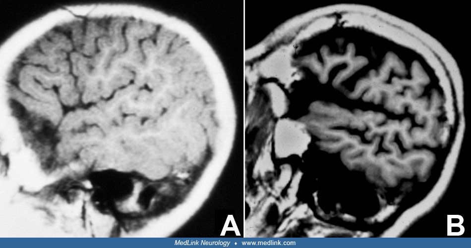
(A) Control. (B) Pontocerebellar hypoplasia type 2. (C) Pontocerebellar hypoplasia type 4. In (A) normal folial development is shown by normal length and 8 to 15 folial branches for each folium. The confines of 1 folium are indicated by a broken line. The dentate nucleus (DN) is shown beneath the cerebellar cortex as an undulating unbroken line. (B) is an example of pontocerebellar hypoplasia type 2 with short folia and limited branching. Space between the folia is depleted (arrows), and only remnants of lost cortex can be found by microscopy in these places. The dentate nucleus (DN) is broken in small islands, possibly resulting from the loss of segments of overlying cortex by antegrade degeneration. (C) shows the findings in cerebellar cortex and dentate nucleus (DN) in pontocerebellar hypoplasia type 4. The cortex is completely denuded, and a ghost folium can only be discerned by its external shape. The gliotic cortex contains microscopic remnants of neurons. Only minute islands of dentate nucleus (DN) remain. (Contributed by Dr. Peter Barth.)