General Neurology
Metal neurotoxicity
Nov. 03, 2025
MedLink, LLC
3525 Del Mar Heights Rd, Ste 304
San Diego, CA 92130-2122
Toll Free (U.S. + Canada): 800-452-2400
US Number: +1-619-640-4660
Support: service@medlink.com
Editor: editor@medlink.com
ISSN: 2831-9125
Toll Free (U.S. + Canada): 800-452-2400
US Number: +1-619-640-4660
Support: service@medlink.com
Editor: editor@medlink.com
ISSN: 2831-9125
Nearly 3,000 illustrations, including video clips of neurologic disorders.
Every article is reviewed by our esteemed Editorial Board for accuracy and currency.
Full spectrum of neurology in 1,200 comprehensive articles.
Listen to MedLink on the go with Audio versions of each article.
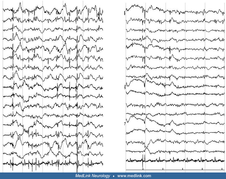
(A) Archimedean spiral: drawing is invalidated by action myoclonus. (B) Somatosensory evoked potentials: stimulation of the left median nerve evokes a typical giant wave at the cortical (N33) level; Averaged polygraph: at the averaged EEG a C4 cortical spike is observed y-time before the myoclonus at the L-biceps level. (C) Sagittal midline T1 brain MRI, slight cerebellar atrophy with mild enlargement of the posterior fossa (white arrow) in an EPM1 patient. (D) Gel electrophoresis showing two bands of 3.6 Kb (indicative of the dodecamer expansion repeat in both mutated alleles) in the homozygous EPM1 patient, as opposed to the native 2.4 Kb bands in the normal and heterozygous carrier individuals. (Contributed by Dr. Pasquale Striano.)