Neuropharmacology & Neurotherapeutics
Rotigotine
Jul. 29, 2021
MedLink, LLC
3525 Del Mar Heights Rd, Ste 304
San Diego, CA 92130-2122
Toll Free (U.S. + Canada): 800-452-2400
US Number: +1-619-640-4660
Support: service@medlink.com
Editor: editor@medlink.com
ISSN: 2831-9125
Toll Free (U.S. + Canada): 800-452-2400
US Number: +1-619-640-4660
Support: service@medlink.com
Editor: editor@medlink.com
ISSN: 2831-9125
Nearly 3,000 illustrations, including video clips of neurologic disorders.
Every article is reviewed by our esteemed Editorial Board for accuracy and currency.
Full spectrum of neurology in 1,200 comprehensive articles.
Listen to MedLink on the go with Audio versions of each article.
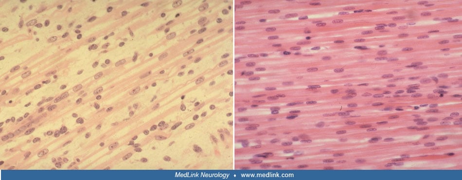
Longitudinal sections of quadriceps femoris muscle of a 14-week human fetus (left) and a term neonate with X-linked myotubular myopathy (right). In both true fetal myotubes and the centronuclear fibers of myotubular myopathy, the nuclei occupy a central portion of the fiber with mitochondria and other organelles in the internuclear spaces. Nuclear chromatin of myotubes is less clumped and more finely dispersed. In fetal myotubes, the distance between nuclei within the same fiber is highly variable; in myotubular myopathy the spacing of nuclei is much more regular. (Used with permission. Sarnat HB. Myotubular myopathy: arrest of morphogenesis of myofibers associated with persistence of fetal vimentin and desmin. Four cases compared with fetal and neonatal muscle. Can J Neurol Sci 1990;17:109-23.)