


Sleep Disorders
Sleep-related leg cramps
Jul. 03, 2026
MedLink, LLC
3525 Del Mar Heights Rd, Ste 304
San Diego, CA 92130-2122
Toll Free (U.S. + Canada): 800-452-2400
US Number: +1-619-640-4660
Support: service@medlink.com
Editor: editor@medlink.com
ISSN: 2831-9125
Toll Free (U.S. + Canada): 800-452-2400
US Number: +1-619-640-4660
Support: service@medlink.com
Editor: editor@medlink.com
ISSN: 2831-9125
Worddefinition
At vero eos et accusamus et iusto odio dignissimos ducimus qui blanditiis praesentium voluptatum deleniti atque corrupti quos dolores et quas.
Corticobasal degeneration is one of the atypical parkinsonian syndromes or parkinsonism-plus syndromes most often characterized by parkinsonism (typically unilateral); apraxia; cortical sensory signs; dystonia; involuntary movements, such as myoclonus; and “alien limb” sign. In this article, the author reviews the clinical features of the disease as well as the pathological findings. Although there is no cure and treatment remains symptomatic, accurate diagnosis can help with patient and family counseling and guide the treating physicians.
|
• Corticobasal degeneration, which is considered as an atypical parkinsonian syndrome or parkinsonism-plus syndrome, is a tauopathy. | |
|
• The pathological hallmark is the deposition of abnormally hyper-phosphorylated microtubule associated protein tau in various parts of the brain. The location of abnormal tau deposition accounts for its heterogeneous presentation. | |
|
• The most common phenotype or presentation of corticobasal degeneration is corticobasal syndrome, which is characterized by parkinsonism with accompanying apraxia, dystonia, myoclonus, and alien-limb phenomenon. | |
|
• Corticobasal syndrome may be seen in other clinically pathologically overlapping tauopathies, such as progressive supranuclear palsy. Features at presentation may aid in distinguishing between the tauopathies. | |
|
• Currently, no specific treatment is available for corticobasal degeneration. |
Corticobasal degeneration was first described by Rebeiz and colleagues via a case series of three patients with progressive asymmetrical parkinsonism and myoclonus (79). In this original description, the term “corticodentatonigral degeneration” was used to describe the pathological changes observed. The disorder was not further described until 1989 by Gibb and colleagues, when the term “corticobasal degeneration” was introduced (28). In the subsequent years, the terms “corticobasal degeneration” and “corticobasal syndrome” have largely been used interchangeably, lending to confusion. To clarify, corticobasal degeneration is the term used to denote the atypical parkinsonian syndrome or parkinsonism-plus syndrome with corresponding pathology demonstrating a specific 4-repeat tauopathy, and corticobasal syndrome is the most common phenotype.
|
• Onset of disease is typically 50 years old or older. | |
|
• Generally, the disease course is gradually progressive over 5 to 7 years. | |
|
• A comprehensive review of pathologically confirmed corticobasal degeneration identified five clinical phenotypes. The most common clinical phenotypes, probable and possible corticobasal syndrome, are characterized by asymmetric parkinsonism (rigidity, dystonia), apraxia, and alien limb phenomena. |
Corticobasal degeneration is a neurodegenerative disorder within the category of tauopathies, a heterogenous group of dementias and movement disorders unified by neuropathological evidence of intracellular filament accumulation composed of microtubule-associated tau protein (51; 57; 12). As a result of differences in where the abnormal tau accumulates in the brain, the clinical presentation of corticobasal degeneration is variable, with corticobasal syndrome the most common phenotype (15).
The onset of disease of corticobasal degeneration is typically 50 years old or older, and the disease course is generally progressive over 5 to 7 years (03), though rapidly progressive cases have been described (53).
The most common phenotype, corticobasal syndrome, is characterized by asymmetrical parkinsonism, dystonia, apraxia, cortical sensory deficits, and alien limb phenomena (cortical signs). Other tauopathies, such as progressive supranuclear palsy, can present with the phenotype of corticobasal syndrome. Apraxia is often a notable feature in the phenotype, corticobasal syndrome.
Revised clinical diagnostic criteria by Armstrong and colleagues identify clinical characteristics that can aid in the diagnosis and distinction from other parkinsonian and dementia disorders (03). In their review of 267 pathologically confirmed corticobasal degeneration cases from published reports and brain banks, clinical corticobasal degeneration phenotypes and diagnostic features were identified. The most common clinical features noted anytime during the clinical course were limb rigidity (85%), bradykinesia or clumsy limb (76%), postural instability (78%), falls (75%), abnormal gait (73%), and axial rigidity (69%). Less commonly seen, but present, were tremor (39%), limb dystonia (38%), and myoclonus (27%). Higher cortical features present in more than five cases included general cognitive impairment (70%), behavioral changes (55%), limb apraxia (57%), aphasia (52%), depression (51%), cortical sensory loss (27%), and alien limb (30%).
In developing corticobasal degeneration diagnostic criteria, five potential phenotypes were described: (1) probable corticobasal syndrome; (2) possible corticobasal syndrome; (3) frontal behavioral-spatial syndrome; (4) nonfluent/agrammatic variant of primary progressive aphasia; (5) progressive supranuclear palsy syndrome (03). The first two phenotypes are the most common. The presence of at least two of the following features would move the diagnosis to probable over possible: asymmetric rigidity, limb dystonia, myoclonus, apraxia, cortical sensory deficit, alien limb phenomena. The frontal behavioral-spatial phenotype is more characterized by the presence of cognitive impairment in the domains of executive function and visuospatial orientation, perhaps with personality changes. The nonfluent variant is characterized agrammatic speech. Lastly, the progressive supranuclear palsy phenotype may be clinically indistinguishable from the other tauopathy, progressive supranuclear palsy, with perhaps impairment in vertical gaze or other eye movement abnormalities, postural instability, axial rigidity, or behavioral changes.
The nonfluent variant of corticobasal degeneration can be one of the etiologies behind a primary progressive aphasia presentation.
There are three types of primary progressive aphasia: (1) progressive nonfluent aphasia, characterized by effortful speech with agrammatism and speech apraxia; (2) semantic primary progressive aphasia, which involves fluent speech with loss of word and object meaning; and (3) logopenic progressive aphasia, characterized by word-finding pauses, moderate anomia, and impaired repetition of sentences (64). In a study of 10 patients with primary progressive aphasia who were followed prospectively until they became nonfluent or mute, Kertesz and Munoz found that all had evidence of frontotemporal dementia at autopsy, but four also had features of corticobasal degeneration (43). Subsequently, published reports have highlighted the emergence of corticobasal degeneration (07; 44) or a mixed pathology in patients who initially present with primary progressive aphasia (19). A longitudinal study compared the clinical and neuroimaging characteristics of patients with nonfluent/agrammatic primary progressive aphasia who were later autopsy-proven to have underlying progressive supranuclear palsy or corticobasal degeneration (89). Other abnormalities of speech in corticobasal degeneration include orobuccal apraxia and a flat, aprosodic speech. Some patients can present with a foreign accent (56). A variety of speech and language abnormalities have been described in patients with corticobasal degeneration (75).
As observed in the most common phenotypes of corticobasal degeneration, probable and possible corticobasal syndrome, the most affected limb can be so dysfunctional that patients find they have little control as it spontaneously floats upward or moves as if no longer attached to the patient's body ("alien limb" phenomenon) (22; 29). Alien limb can be seen as a motor or sensory-based phenomenon. Although the alien hand phenomenon is common (observed in 50% to 60% patients), rarely, patients may also develop alien leg phenomenon (29; 70). In a cohort of 150 patients with alien limb phenomenon studied by Graff-Radford and colleagues, 108 patients had a diagnosis of corticobasal degeneration (29).
At times, the presence of cognitive impairment may bring an individual with corticobasal degeneration to the physician’s attention, as opposed to the motor features that may or may not be present. In a series of autopsy-proven cases of corticobasal degeneration, dementia was the most frequent clinical presentation (33). On neuropsychological testing, the cognitive profile of those with corticobasal degeneration appears similar to that observed with other tauopathies, such as progressive supranuclear palsy or frontotemporal dementia, but language may be disproportionately impacted in those with the nonfluent variant. In general, the cognitive profile observed in corticobasal degeneration is typically a dysexecutive syndrome (77; 31). Disinhibition, perseveration, and hemispatial neglect are alternate neuropsychological presentations (46). Semantic memory deficits are particularly marked for numbers (34). Cortical sensory deficits, unusual in most parkinsonian syndromes, can occur in approximately one third of patients and can be the presenting symptom (83; 25; 30; 84). Approximately 15% of patients develop dysphasic signs (82; 83; 41). Cases of corticobasal degeneration presenting with other cortical findings, such as progressive apraxic agraphia or acalculia, have also been described (71; 73; 69).
Clinical features at presentation and disease course were examined in patients with pathologically, genetically, and biochemically verified corticobasal degeneration and in those with other tauopathies with clinical features of corticobasal syndrome (02). The authors found that clinical features of “freezing” of gait at onset or absence of dysarthria at presentation in setting of age at onset under 66 years were predicators of corticobasal degeneration pathology (02).
Corticobasal degeneration is a gradually progressive neurodegenerative disease that is without a cure at this time. The onset of disease of corticobasal degeneration is typically 50 years old or older, and the disease course is generally progressive over 5 to 7 years (03), though rapidly progressive cases have been described (53). The progression is typically faster than that of typical idiopathic Parkinson disease. Ling and colleagues described cases of corticobasal degeneration with an average disease duration of 3 years or less (53). The degree of tau burden in the brains of those impacted seemingly corresponded to the rapidity of disease progression. Advanced disease was marked by deterioration in motor and cognitive function, with increasing falls, dysphagia, dysarthria with increasing risk for aspiration, incontinence, and weight loss. In a series of pathologically proven cases, dysarthria occurred 40 months after disease onset and dysphagia at a median of 64 months, in contrast to 84 months and 130 months in Parkinson disease (65). At this time, there are no disease-modifying therapies, meaning medications do not appear to affect the natural progression of the neurodegenerative process, and the disease usually progresses to death.
A 57-year-old librarian described an 8-month history of left-sided hand and leg incoordination. Specifically dating the onset of difficulty was challenging, but she experienced increasing slowness and loss of agility in the left hand when she performed tasks such as dressing, sorting cards, or typing. Often, the left hand cramped, and over the previous month she noted that the whole hand or index fingers occasionally moved by themselves. The left leg also cramped and inverted after she walked more than 200 feet. She felt her right side was unaffected, and her thinking was normal, although she was easily startled. She experienced general body jerks when reading quietly at night. She stumbled but had no falls.
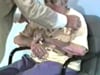
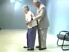
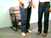
Clinical findings. The patient had a masked face and slight impairment of vertical conjugate gaze. She had parkinsonian signs of rigidity and bradykinesia bilaterally; they were more severe on the left. The left hand and foot had dystonic posture. When the patient closed her eyes and outstretched her hands, the left hand drifted upward, and the fingers wiggled; "They've a mind of their own," she commented. There were intermittent myoclonic jerks of all extremities. Primary sensory modalities were normal, but she had astereognosis in the left hand and could not imitate hammering or saluting to a verbal command with the left hand. Her gait was shuffling, and to a postural threat, she took three steps backwards.
Diagnostic tests. Lab tests, including thyroid functions, were normal. MR scan showed cerebral atrophy, particularly of the right parietal region. SPECT scan showed asymmetric uptake of DaTSCAN, especially in the right basal ganglia.
Clinical course. She received carbidopa and levodopa at 25 and 100 mg three times per day; there were no signs of benefit. Dopamine agonists only resulted in nausea and dizziness secondary to hypotension. The hand cramp became more troublesome, and she received focal botulinum toxin injections, with relief of pain and spasm. Gait deteriorated with increased falls, and she eventually became wheelchair dependent. The right upper extremity became markedly bradykinetic and rigid in a flexed position. Myoclonus increased and was rhythmic at times, but no seizures developed. Clonazepam abated the myoclonus, but only low doses were used because of sedation. The patient died 5 years after onset.
Corticobasal degeneration falls into a category of diseases called tauopathies (35). Corticobasal degeneration is predominantly a sporadic disease, but familiar cases due to microtuble associated tau protein (MAPT) mutations have been reported (92). Familiar cases linked to specific genetic mutations provide insight into pathology, even in sporadic forms. Tau is a protein that was originally implicated in disease in 1997 (91), followed by the discovery of mutations in the tau gene in frontotemporal dementia and parkinsonism linked to chromosome 17 (FTDP-17). Subsequently, tau pathology was implicated in other pathologically similar diseases, such as Alzheimer disease, Pick disease, and progressive supranuclear palsy (82; 39). The pathological process can involve different tau isoforms such as 3R-tau, 4R-tau, or mixed 3R-/4R-tau isoforms. Corticobasal degeneration has been recognized as a 4R-taupathy (35). Familial cases of corticobasal degeneration or families with both corticobasal degeneration and frontotemporal dementia have been linked to mutations of chromosome 17 (86). In addition, although the microtuble-associated protein tau gene (MAPT) has been associated with autosomal dominant forms of dementia, it may be a factor in sporadic corticobasal degeneration. A case of a 41-year-old gentleman with the corticobasal degeneration phenotype was found to have G389R mutation in the MAPT gene (85). The clinical features of corticobasal degeneration, including primary progressive aphasia, have been described in two cases with LRRK2 G2019S mutation (13).
A kindred with some members having clinically suspected corticobasal degeneration and others with frontotemporal dementia suggest that common underlying etiologies may have different clinical phenotypes (10). A genome-wide association study reported that corticobasal degeneration and progressive supranuclear palsy share a common genetic risk factor other than MAPT at 3p22 MOBP (myelin-associated oligodendrocyte basic protein) (48).
Other genetic etiologies continue to be explored with the use of whole genome sequencing, but no etiology has yet been determined. Pathologically, corticobasal degeneration has distinctive features that include prominent frontoparietal cortical atrophy and degeneration of the substantia nigra pars compacta. When atrophy is diffuse, other diseases must be considered, such as frontotemporal lobar degeneration or Alzheimer disease, but focal atrophy suggests corticobasal degeneration (100). Although the clinical presentation is usually asymmetric, pathological changes and cortical atrophy at death may be symmetric or asymmetric with typically prominent atrophy of the precentral gyrus (16). Microscopically, the traditional hallmark sign is the presence of swollen achromatic cells (ballooned neurons) that resemble those seen in Pick disease (80; 28). The presence of tau pathology in astrocytes is now considered to be pathognomonic for corticobasal degeneration (15). A set of minimal neuropathologic consensus criteria was established in 2002 for corticobasal degeneration by the Office of Rare Diseases. These are: cortical and striatal tau-positive neuronal and glial lesions, especially astrocytic plaques and thread-like lesions in both gray and white matter, as well as neuronal loss in focal cortical regions and in the substantia nigra (21). Corticobasal degeneration can be distinguished pathologically from the other tauopathies, progressive supranuclear palsy, and frontotemporal dementia, but variants of frontotemporal dementia may be more difficult to distinguish (40).
Studies of tau protein demonstrate that corticobasal degeneration shares a common genetic background with progressive supranuclear palsy (04). Both share the same tau H1 haplotype (36). This finding suggests a similar genomic cause and introduces the concept that these two distinct conditions may be phenotypically distinct prototypes of a single biological disorder.
Given the pathology, it is not surprising that with further use of genetics in corticobasal degeneration that tau associated genes are being discovered as potential underlying etiologies of the disease. More novel mutations seem to be identified as time and more samples are studied (01).
Several experimental approaches in animal models have attempted to reduce the tau-burden in neurodegeneration (90; 60).
Studies on pathophysiology have included analysis of cortical disinhibition with transcranial magnetic stimulation (26). In one review, neurophysiological abnormalities in corticobasal degeneration are explained in detail (67).
Some cases suggest a possible role of the TAR-DNA-binding protein 43 (TDP-43). This nuclear protein is typically involved in transcriptional repression and splicing but is found in inclusions in a range of neurodegenerative disease including amyotrophic lateral sclerosis, Alzheimer disease, and frontotemporal lobar degeneration (99). It has been found in over 15% of corticobasal degeneration cases (96). Another potential etiology is a mutation along the same pathway that results in abnormal tau phosphorylation, specifically progranulin mutation (93; 94).
The rarity of the disease makes epidemiological studies difficult to perform. Disease registries have been used to estimate the prevalence of corticobasal degeneration (95). It has been reported to be 10 times rarer than a clinicopathologically similar tauopathy, progressive supranuclear palsy (57). There is a mild predominance of men. No geographic predominance or ethnic clustering has been reported.
No information is known of preventing corticobasal degeneration.
Although early reports suggested that corticobasal degeneration was a clinically distinct disorder, additional studies have shown that the most common phenotype, corticobasal syndrome, can develop in the context of other histopathological diagnoses like progressive supranuclear palsy, Pick disease, Creutzfeldt-Jakob disease, Alzheimer disease, progressive multifocal leukoencephalopathy, Fahr disease, motor neuron disease-inclusion dementia, neurosyphilis, and sporadic spinocerebellar ataxia type 8 (09; 32; 97; 50; 55; 08; 05). Conversely, cognitive impairment and supranuclear gaze palsies that may suggest other parkinsonism-plus syndromes can occur in corticobasal degeneration (81). Ocular problems are most suggestive of progressive supranuclear palsy, whereas pyramidal signs, weakness, and Babinski signs, when apparent, may suggest multiple system atrophy. If dementia predominates, the picture might suggest focal Alzheimer disease or frontotemporal dementia. Pick disease can present with all the typical features of corticobasal degeneration including parkinsonism, myoclonus, and asymmetric dyspraxia (52; 63). When myoclonus predominates and the disease course is rapidly progressive, Creutzfeldt-Jakob disease or other disorders with secondary myoclonus should be considered (58; 45). Other non-Alzheimer dementias, such as dementia with Lewy bodies, primary progressive aphasia, semantic dementia, frontotemporal dementia, and familial forms of frontal lobe dementia, must also be considered in cases where cognitive decline predominates (11; 72; 42; 62; 34). MR-based volumetry models have been developed to help differentiate progressive supranuclear palsy from corticobasal degeneration (87).
Although apraxia, alien limb, or arm levitation are often hallmarks of corticobasal degeneration, such findings can occur in progressive nuclear palsy, Alzheimer disease, Pick disease, and Creutzfeldt-Jakob disease (06; 09). In comparison with progressive supranuclear palsy, however, apraxic errors in corticobasal degeneration are more frequent and severe, involving both transitive and intransitive tasks (76).
The diagnosis of corticobasal degeneration is primarily a clinical one based on history and examination. However, studies such as the DaTscan can be used as supportive studies, though the DaTscan cannot distinguish between neurodegenerative parkinsonian disorders. Advances in volumetric MR imaging and paired disease progression modeling are being made to aid in diagnosis. Saito and colleagues demonstrated that differences in temporal progression patterns of brain atrophy utilizing SuStain can aid in the distinction between corticobasal degeneration and progressive supranuclear palsy (87). CT or MR imaging may show signs of generalized cerebral or focal parasagittal and parietal atrophy. Generally, CT or MR imaging is performed to rule out a structural lesion, such as hydrocephalus, stroke, or mass, that may account for presentation. Labs to look for other potentially reversible contributors, such as syphilis or Lyme testing, thyroid studies, and vitamin levels (most commonly vitamin B12 and folate), are generally done. Cerebrospinal fluid analysis may be done to rule out other conditions, such as prion disease, but largely remains a research tool. CSF studies may help in the future for differentiating corticobasal degeneration from other conditions.
Asymmetry of metabolic activity in the parietal lobe contralateral to limb apraxia can also be documented by PET scans (24; 23; 38). Hypometabolism of the superior parietal lobule and supplementary motor cortex among subjects with visuoimitative upper limb apraxia in corticobasal degeneration further suggests direct involvement of parietofrontal neural networks (74).
DaTscan or DAT-SPECT utilizes dopamine transporter imaging as a functional measure. In a retrospective analysis of dopamine transporter (DAT) SPECT studies in two patients with a pathologically confirmed corticobasal syndrome, baseline scans at 1.5 years after symptom onset revealed only mild abnormalities (reduced uptake in one putamen). Follow-up scans at 4.5 years (case 1) and 5 years (case 2) after symptom onset showed a marked decline of striatal DAT binding. In both cases, there was a 37% binding reduction from the age-expected striatal binding value. Therefore, patients with corticobasal degeneration may have delayed neuronal loss in the substantia nigra and follow-up DAT imaging may be of value in patients with possible corticobasal degeneration and a normal baseline scan (78). In a study on 34 patients with corticobasal degeneration, using FDG-PET and DAT-SPECT, Mille and colleagues revealed asymmetric cortical and subcortical hypometabolism, symmetrically reduced metabolism in the thalamus, and slightly asymmetric reduction in DAT (61).
In utilizing neuropsychological testing, patterns of apraxia deficit may help differentiate cases of corticobasal degeneration from those with progressive supranuclear palsy (20). Another four pathologically confirmed cases suggested that early stage corticobasal degeneration may present with self-centered behaviors, such as frontotemporal dementia, apathy with and without auditory hallucination, and aggressive behaviors with delusion and visual hallucination prior to the onset of motor manifestations (37).
Studies utilizing MRI brain imaging findings, namely patterns in areas of atrophy and subcortical white matter hyperintensities, are demonstrating distinguishing features in those with pathologically confirmed corticobasal degeneration distinct from those with Alzheimer disease, dementia with Lewy bodies, progressive supranuclear palsy, and frontotemporal lobar degeneration (88).
At this time, there is no cure, and the therapeutic approach is aimed at addressing symptoms. Levodopa is the most effective agent for the parkinsonian motor features (47). In a retrospective study on pathologically proven cases of corticobasal degeneration, levodopa had resulted in mild-to-moderate improvement (54). Side effects of levodopa, like dyskinesia, are not typical in corticobasal degeneration but can occur (27). Levodopa is generally less well-tolerated in the atypical parkinsonian disorders, like corticobasal degeneration, than in idiopathic Parkinson disease (98).
For myoclonus, levetiracetam and clonazepam have been reported to be beneficial (14; 49). Major side effects of these medications include changes in mental status and mood. For the symptomatic treatment of dystonia, botulinum toxin may be helpful (47; 17; 66). Mood disorders like depression and anxiety are common, and selective serotonin reuptake inhibitors (SSRIs) are effective (59) and generally better tolerated than tricyclic antidepressants, which should be avoided in view of their disabling anticholinergic side effects. Antipsychotics, other than quetiapine and clozapine, are contraindicated given their dopamine-blocking properties.
Several therapeutic approaches targeting the potential underlying biological processes are in clinical trials to reduce the progression of the pathology. These approaches include reduction of abnormal post-translational modifications, blocking of transcellular spread of tau, stabilization of microtubules and neuroprotection, and enhancement of tau degradation (68). At the time of this update, there are currently four therapies, one mAB and three small molecules, in phase III trials and 16 drugs in phase II to potentially target tauopathies (18).
There are excellent reviews of pharmacological, diet, and other symptomatic therapies for corticobasal degeneration (49; 59).
Unknown, but generally not applicable. Age of disease onset is usually among postmenopausal women.
Delirium is common among postoperative older adults, those with dementia, and those with neurodegenerative parkinsonian disorders. Patients with neurodegenerative parkinsonian disorders should not be exposed to dopamine-blocking agents, such as antipsychotics or some antiemetics.
All contributors' financial relationships have been reviewed and mitigated to ensure that this and every other article is free from commercial bias.

Katherine Amodeo MD
Dr. Amodeo of School of Medicine at New York Medical College has no relevant financial relationships to disclose.
See Profile
Robert Fekete MD
Dr. Fekete of New York Medical College received consultation fees from Acadia Pharmaceutical, Acorda, Adamas/Supernus Pharmaceuticals, Amneal/Impax, Kyowa Kirin, Lundbeck Inc., Neurocrine Inc., and Teva Pharmaceutical, Inc.
See ProfileNearly 3,000 illustrations, including video clips of neurologic disorders.
Every article is reviewed by our esteemed Editorial Board for accuracy and currency.
Full spectrum of neurology in 1,200 comprehensive articles.
Listen to MedLink on the go with Audio versions of each article.
MedLink, LLC
3525 Del Mar Heights Rd, Ste 304
San Diego, CA 92130-2122
Toll Free (U.S. + Canada): 800-452-2400
US Number: +1-619-640-4660
Support: service@medlink.com
Editor: editor@medlink.com
ISSN: 2831-9125
Sleep Disorders
Jul. 03, 2026
Neurobehavioral & Cognitive Disorders
Jun. 17, 2026
Movement Disorders
May. 24, 2026
Neurobehavioral & Cognitive Disorders
May. 20, 2026
Movement Disorders
May. 18, 2026
Movement Disorders
May. 18, 2026
Movement Disorders
May. 18, 2026
Movement Disorders
May. 18, 2026