


Epilepsy & Seizures
Driving and epilepsy
Jul. 08, 2026
MedLink, LLC
3525 Del Mar Heights Rd, Ste 304
San Diego, CA 92130-2122
Toll Free (U.S. + Canada): 800-452-2400
US Number: +1-619-640-4660
Support: service@medlink.com
Editor: editor@medlink.com
ISSN: 2831-9125
Toll Free (U.S. + Canada): 800-452-2400
US Number: +1-619-640-4660
Support: service@medlink.com
Editor: editor@medlink.com
ISSN: 2831-9125
Worddefinition
At vero eos et accusamus et iusto odio dignissimos ducimus qui blanditiis praesentium voluptatum deleniti atque corrupti quos dolores et quas.
The electroencephalogram (EEG) is a widely available, cost-effective, portable neurophysiological study of brain function with worldwide applications. As a window into the brain, it remains the foundational diagnostic tool to evaluate people suspected of seizures. It is a safe, noninvasive, diagnostic, 2-dimensional clinical study used to assess the brain’s 3-dimensional electrophysiological activity. EEG is the most useful test for evaluating people with possible epilepsy when epileptiform activity is demonstrated. It may provide specific neurophysiological information to support clinical diagnoses in the EEG laboratory, epilepsy monitoring unit, emergency room, inpatient wards, including the intensive care unit, or operating room and can assist in the evaluation of people in an ambulatory setting. When interictal epileptiform discharges are recorded on EEG, it serves to classify the seizure type or epilepsy syndrome that is suspected based on clinical grounds. Recent seizure classification systems have been developed to subdivide them into focal, generalized, unknown (whether focal or generalized), and unclassified onset. Being aware of seizure and epilepsy classification is essential to guide selection of the most appropriate antiseizure medication across the lifespan (67; 09). EEG is an important adjunct to the clinical examination for diagnosing and treating epilepsy as well as subclinical or unrecognized seizures. It is foundational in the diagnosis of nonconvulsive status epilepticus in critically ill patients when the clinical examination is unrevealing. Clinical diagnostic and management algorithms for EEG and for epilepsy have been specifically developed to minimize misdiagnosis and mistreatment of patients with seizures. Continuous EEG in the ICU has devised validated terminology for EEG increasingly used to identify and treat patients with electrographic seizures and status epilepticus (18; 41). Rapid EEG systems are becoming widely used in urgent evaluation of status epilepticus (21; 33). In the operating room, electrocorticography helps neurosurgeons define surgical borders of functional and epileptogenic tissues (31). Chronic electrocorticography obtained from a responsive neurostimulation device assists in treatment decision-making. With the increasing emphasis on outpatient management, ambulatory EEG has become a staple in assessing patients with home video EEG telemetry to record a patient’s EEG in the home environment (37).
Overall, the utility of EEG has evolved into a sophisticated computer-based clinical and research tool that is fundamental for exploring essential brain functions (104). Despite the innovation of neuroimaging the anatomy of the brain, EEG continues to have a vital role in the dynamic physiological evaluation of neurologic disease. In this way, it extends usage in patients with seizures and epilepsy to diagnosis and monitoring neurologic disorders, such as encephalopathy, traumatic brain injury, sleep disorders, coma, and brain death. It is incumbent on the EEG interpreter to know how to identify the essential characteristics to report accurate impressions for clinical use (91).
|
• EEG is the diagnostic test of choice when evaluating a patient with seizures and epilepsy, though pitfalls exist in interpretation because of artifacts, normal variations, and benign variants that mimic epileptiform abnormalities. | |
|
• Epilepsy is a clinical diagnosis that is supported and classified by epileptiform activity on the EEG and confirmed when a seizure is recorded in a patient. | |
|
• Misdiagnosis of nonepileptic events as seizures is not a rare situation due in part to EEG misinterpretation. | |
|
• Long-term video EEG monitoring in the epilepsy monitoring unit and continuous EEG in the intensive care unit may clarify the seizure burden for treatment. | |
|
• The surgical treatment of epilepsy relies on the interictal and ictal EEG to characterize the electroclinical localization of the epileptogenic zone. | |
|
• The sophistication of EEG has expanded with the use of automation and implementation of artificial intelligence (AI) to aid in diagnostic evaluation of patients with epilepsy and other brain diseases. |
In his seminal work “Uber das Elektroenkephalogram des Menschen” (“On the EEG of Man”), Hans Berger pioneered the discovery of the human EEG, first recorded in 1929 (36). The practical usefulness of EEG became apparent in the 1930s after interictal discharges were demonstrated first by Fisher and Lowenback and later by Gibbs, David, and Lennox in the United States.
In 1936, W Gray Walter demonstrated that this technology could aid in the diagnosis of tumors, stroke, and other focal brain disorders. For 40 years, EEG was the cornerstone to the diagnosis and treatment of seizures and epilepsy. Until the advent of CT and MRI, it was the first-line neurodiagnostic test for diagnosing tumors, stroke, and other focal brain disorders.
EEG data were analyzed by an orderly visual approach to interpretation until the 1960s, when digital equipment was introduced to begin the digital age. In the 1970s, the Fourier transform, computer-based algorithms and analytics, quantitative EEG, and trend analysis became a reality. Recent advances have centered on transforming discrete sources as stable linear dipoles to model EEG using inverse methods to depict the EEG in source space using MRI co-registration. Furthermore, newer AI models use algorithms capable of accurately “reading” EEG to differentiate normal from abnormal recordings as well as classifying focal and generalized slowing and epileptiform discharges.
Terminology. In 2017, after 35 years, the International League Against Epilepsy (ILAE) released a new classification of seizure types. This was largely based on the existing classification originally formulated in 1981 (28). Imminent classification systems are now in progress to further refine standardized terminology for seizure classification (09). In broad terms, epilepsy syndromes are classified as generalized, focal, unknown, and unclassified with an etiology that is genetic, structural-metabolic, or unknown (28; 09). Seizures are classified according to where they start in the brain (97; 91). Generalized seizures originate within both hemispheres and rapidly engage bilaterally distributed networks of neurons at onset (44). The term “bilateral” is used for focal seizures that propagate to both hemispheres (ie, focal to bilateral tonic-clonic seizures), and “generalized” is a term used for seizures that begin simultaneously in both hemispheres.
Focal seizures refer to those seizures that originate within networks limited to one hemisphere. Focal seizures may start on the surface of the brain or arise in deeper structures and remain restricted to an area within the hemisphere (focal seizures with preserved consciousness) or spread beyond a single hemisphere to involve a larger network (focal seizures with impaired consciousness) and focal to bilateral tonic-clonic seizures. Focal seizures are also classified as with and without observable motor manifestations. Semiology descriptors involving signs and symptoms are provided in chronological order. Unknown seizures are those that cannot be defined as focal or generalized.
The application of the standard scalp EEG in epilepsy has relied on the electrocerebral activity largely between the 1 to 30 Hz bandwidth (ie, Berger’s bandwidth). Filter settings are increasingly being “opened” during video epilepsy monitoring to obtain full-band EEG, providing a more comprehensive approach (96). Newer applications of high frequencies (ie, gamma and high-frequency oscillations) may disclose the brain regions and networks that are involved in seizure genesis (Table 1). Faster frequencies are particularly important when using invasive EEG techniques as they may not always be visible on qualitative assessment using scalp EEG for recording.
|
Frequency (Hz) |
Bandwidth |
Normal |
Pathological |
|
0.0 - 0.5 |
Infraslow activity* |
Artifacts |
Onset of focal seizures |
|
0.5 - 3.5 |
Delta |
Sleep, HV, PSWY, elderly |
Encephalopathy, white matter lesion |
|
>3.5 - <8.0 |
Theta |
Drowsiness, children, elderly |
Encephalopathy, white matter lesion |
|
8 - 13 |
Alpha |
PDR, mu rhythm, “third” rhythm |
Ictal rhythm in seizure, alpha coma |
|
13 - 30 |
Beta |
Medication, drowsiness |
Breach rhythm, drug overdose, ictal rhythm |
|
30 - 80 |
Gamma* |
Voluntary motor movement, learning/memory |
Seizures |
|
80 - 250 |
Ripples* |
Cognitive processing/memory |
Interictal and ictal seizure frequency, possible epileptogenesis |
|
250 - 500 |
Fast ripples* |
? |
Focal seizures |
|
500 - 1000 |
Very fast ripples* |
Acquisition of sensory information |
Seizures |
|
| |||
|
HV=hyperventilation; PSWY=posterior slow waves of youth; PDR=posterior dominant rhythm | |||
The EEG is composed of a combination of frequencies to be visualized as a complex mix of waveforms. Characteristic interictal EEG features of epilepsy are spikes (20 to 70 msec) and sharp waves (70 to 200 msec) when displayed on a review monitor at a display speed of 30 mm/second. True epileptiform discharges are distinguished from other sharply contoured waveforms by appearing dissimilar to the background waveforms surrounding them, disrupting the background, occupying a “definable physiological field,” having a rapid rise with an asymmetric appearance, and being associated with an after-going slow wave. The criteria for identifying them require several of these features to be present (47; 58). This also presumes that benign variants and artifacts have been excluded (47). Spikes and sharp waves can present in isolation or as polyspikes or polysharp waves. Spikes and sharp waves possess the same potential for seizure genesis independent of morphology.
Each type of EEG recording in epilepsy has its own advantages and disadvantages. Standard scalp EEG recordings, short-term and rapid EEG, computer-assisted ambulatory EEG (CAA-EEG), and in-patient continuous video EEG monitoring (VEM) are different methods of EEG recording (51), each with different clinical benefits and limitations (Table 3).
Scalp EEG is the simplest, least expensive, most practical, and, therefore, most common method to acquire EEG during routine clinical use. A standard scalp EEG is usually a brief 20- to 30-minute recording (but may extend up to 60 minutes duration). It is, therefore, typically inadequate for capturing infrequent paroxysmal neurologic events and abnormalities (05). A 5-year study of 175 outpatient short-term EEGs (shorter than 24 hours) found that only 7% yielded seizures during the recording (76). A prospective study of EEGs from 1803 patients revealed that only 19% of the patients had interictal epileptiform discharges in the first 30 minutes of recording, but the event capture rate increased by 30% during longer recordings (13).
Prolonged EEG recording using either CAA-EEG or inpatient video EEG monitoring offers distinct advantages. Another prospective study of 100 patients evaluated after a first unprovoked seizure demonstrated that 24 hours of ambulatory EEG recording was significantly more sensitive in detecting interictal discharges and seizures when compared to a first or second shorter routine EEG (72% vs. 11% vs. 22%, respectively) (38). A higher diagnostic yield (40%) was found in children when EEG monitoring was longer than 6 hours (82). The yield of identifying epileptiform discharges to support a clinical diagnosis of epilepsy has been approximately 2.0 to 2.5 times that of a standard EEG and was cost-effective when compared to the gold standard of video EEG monitoring with a yield of ambulatory-EEG greater than 70% (19).
In critically ill patients, studies have demonstrated that though most seizures are captured within 24 hours of recording, some may require even longer monitoring depending on risk factors and clinical history, up to 48 hours (84; 119). On the other hand, routine brief EEG recordings in the hospital can be of value in predicting the risk of seizures, need for continuous EEG monitoring, and institution of anti-seizure medications (85; 50).
The American Clinical Neurophysiology Society has established guidelines for conducting EEG in adults and children (www.acns.org/practice/guidelines). Guidelines for video EEG monitoring and continuous EEG monitoring for seizures in the intensive care unit are also available. The American Board of Registration of Technologists in EEG and Evoked Potentials has devised standards for performing EEG (www.abret.org). The American Board of Clinical Neurophysiology and the American Board of Psychiatry and Neurology have set minimal competency examinations for physicians interpreting EEG. Clinical uses have been further detailed by the American Clinical Neurophysiology Society (ACNS), American Epilepsy Society (AES), and American Academy of Neurology (AAN) to detail the clinical usefulness of EEG in adult and pediatric populations.
Epilepsy is a common and serious neurologic disease. It has a lifetime prevalence of 7.60 per 1000 people. More than 70 million people are affected by epilepsy worldwide. EEG is fundamental in its application for patients suspected of seizures or epilepsy. It can be useful in the following clinical scenarios by leading to an alteration in management or prognosis.
|
• After a first seizure. When epileptiform activity is recorded in the presence of a seizure, the recurrence risk is great enough in most patients (at least 60%), and a diagnosis of epilepsy reached. | |
|
• After a nondiagnostic standard EEG in suspected epilepsy. Repeat or prolonged EEGs will increase the yield of demonstrating an epileptiform abnormality. | |
|
• In the classification of seizure (disorders and syndromes when the diagnosis of focal or generalized), epilepsies are unclear on clinical grounds alone (eg, seizures associated with staring). EEG can contribute to the epilepsy diagnosis and monitor the response to treatment. | |
|
• Prior to tapering antiseizure medications after prolonged seizure-free periods. Epileptiform activity, especially generalized spike-and-slow waves, demonstrates the potential for manifesting seizures independent of historical reporting. | |
|
• Quantification of seizures when epilepsy produces frequent and subtle seizures, such as absence seizures or subtle focal seizures with impaired consciousness.* | |
|
• Behavioral disorders and impaired communication. When patients can't communicate symptoms, EEGs offer objective data on physiological causes at home or in hospital.* | |
|
• Hospital patients who experience acute mental status changes. Patients may struggle to describe seizure episodes, but EEG can detect subclinical seizures.* | |
|
• Characterizing seizure onset during an evaluation of patients for epilepsy surgery.* | |
|
| |
The EEG alone should not be used to exclude a diagnosis of epilepsy or to support the presence of a structural lesion of the brain. MRI or CT brain neuroimaging are more definitive and reliable, especially for subcortical locations in the brain.
Generally, recording a standard scalp EEG is safe. It is a noninvasive test, and rarely, patients have reported allergies and reactions to electrodes or paste/gel. Activating procedures may precipitate lightheadedness, tingling, and rarely, syncope during hyperventilation. There are no contraindications to EEG recording in pregnancy; however, activation procedures are sometimes avoided because of the small risk of inducing a complication such as seizure or inducing preterm labor during pregnancy with adverse influence on the developing fetus. Hyperventilation is breathing deeply at a rate of 18 to 24 breaths per minute for 2 to 3 minutes. Relative contraindications to hyperventilation include the following (56):
|
• Increased intracranial pressure | |
|
• Uncontrolled hypertension | |
|
• Recent stroke, transient ischemic attacks, intracranial hemorrhage, or severe cerebrovascular disease, including carotid artery stenosis and vasospasm | |
|
• Severe cardiac disease, recent myocardial infarction, or angina pectoris | |
|
• Severe pulmonary disease, chronic obstructive pulmonary disease, bronchitis, asthma, or patients requiring oxygen therapy | |
|
• Recent surgery | |
|
• Moyamoya disease | |
|
• Hyperviscosity syndromes and sickle-cell anemia | |
|
• Pregnancy |
Intermittent photic stimulation is routinely performed during EEG. The patient is presented with a series of photic flashes delivered by a strobe light at a measured distance of 20 to 30 cm from the patient’s head. A series of stimulus trains of flashing light are presented for 4 to 10 seconds, with at least 4 seconds between each train successively up to 30 Hz. Intermittent photic stimulation should be performed at a separate time from hyperventilation. Significant ophthalmologic conditions, migraine, and recent surgeries are a relative contraindication. Neither diagnosis of epilepsy nor a historical report of seizures is a contraindication to photic stimulation. However, persistent use of photic stimulation when a photoparoxysmal response has been demonstrated should be avoided given the risk of potentiating a clinical seizure. The photoparoxysmal response occurs when photic stimulation produces bilaterally synchronous epileptiform discharges. The discharges may outlast the stimulus by several seconds (non-self-limited) and may portend a greater likelihood of precipitating a seizure self-limited to the duration of the photic stimulation, which may exist as a genetic trait independent of epilepsy. Patients with idiopathic generalized epilepsies and occipital epilepsies should be made aware of the possibility of a breakthrough seizure during intermittent photic stimulation.
Interictal EEG. EEG features are divided into the following phases:
|
• Interictal – present between clinical seizures |
The utility of interictal EEG has been the foundation for evaluating patients with epilepsy (65; 96; 104). It is rare to capture a seizure during a routine outpatient EEG in adults due to the limited duration of recording; however, interictal epileptiform discharges are very specific and are highly correlated with epilepsy in the proper clinical setting, though they are not diagnostic for epilepsy (54). The prevalence of interictal epileptiform discharges in those with epilepsy depends on a number of factors. It has been reported that 29% to 55% of adults with epilepsy will show one or more interictal epileptiform discharge on a single EEG (120; 72; 74). With repeated testing, including sleep recording, an interictal epileptiform discharge is seen in the EEG of 59% to 92% of epilepsy patients with the fourth recording, but there is minimal additional yield thereafter (120; 72; 11).
The sensitivity of EEG to detect interictal epileptiform discharges varies considerably depending on the age of the patient, presence of intracranial lesion, seizure frequency, epilepsy syndrome, and the site of seizure onset, as well as the duration of EEG recording. The diagnostic yield of the EEG can be increased by sleep deprivation, recording sleep, serial studies, prolonging the recording, and early post-seizure recording.
Interictal epileptiform discharges may also be seen as an inherited trait in patients without a clinical diagnosis and can infrequently be found in asymptomatic and healthy individuals (65). The sample sizes of asymptomatic adults with interictal epileptiform discharges who have follow-up data are small, but it appears that less than 3% go on to have seizures (120; 35; 77). One study of 13,658 adult males aged 17 to 25 without a previous history of significant illness who were medically screened for training in the Royal Air Force of England yielded 69 (0.5%) military personnel with unequivocal interictal epileptiform discharges (35).
Common types of interictal epileptiform discharges seen in asymptomatic individuals include centrotemporal spikes, occipital spikes, and a self-limited photoparoxysmal response (16; 35).
About 2% to 3% of asymptomatic school-aged children have centrotemporal spikes, but only a small fraction of these, possibly as low as 10%, will develop self-limited childhood epilepsy with centrotemporal (Rolandic) spikes with recurrent seizures (16). However, the presence of interictal epileptiform discharges in the EEG of asymptomatic individuals is associated with an increased likelihood of developing epilepsy. In patients with a pathological condition of the brain, the incidence of interictal epileptiform discharges is increased. Patients with interictal epileptiform discharges and congenital brain malformations, brain tumors, prior craniotomy, and intellectual disability are at high risk for subsequently manifesting clinical seizures (120). Some medications may also cause interictal epileptiform discharges. In a series of 3,143 nonepileptic psychiatric patients, interictal epileptiform discharges were found in 3.0% to 3.8% of patients taking major tranquillizers, antidepressants, or lithium compared with 0.8% of patients taking benzodiazepines (77).
Ictal EEG. The ictal EEG reflects an electrographic pattern that is characteristic of patients with epilepsy. It is identified by recurrent epileptiform activity that begins abruptly and continues to evolve in frequency and spatial distribution following a paroxysmal occurrence. The onset and termination are usually abrupt, with changes that take place over seconds to a couple of minutes. Seizures that occur for more than 5 minutes are “prolonged,” and convulsive seizures lasting this duration are diagnostic of status epilepticus (107).
The ictal EEG is generally composed of a rhythmic pattern that increases and decreases in frequency. Temporal evolution occurs, and the ictal activity displays increasing amplitude and often crescendo-decrescendo (focal) or decrescendo (generalized) changes in frequency. There is also spatial evolution that occurs due to seizure propagation as new regions of the brain become involved with evolution to include a broader field of spread. The morphologic characteristics of the ictal EEG can vary by the seizure type. For example, frontal lobe seizures may be marked by a nonlocalized diffuse attenuation or generalized theta/delta but may also occur with lateralized, low-voltage, ictal fast activity. The site of seizure onset and the frequency and topographic distribution of the ictal EEG is usually a better marker. In mesial temporal lobe epilepsy, the ictal scalp EEG is characterized by a rhythmic theta frequency (5 to 9 Hz), whereas in lateral temporal lobe epilepsy, it manifests a rhythmic delta frequency (lower than 4 Hz) at seizure onset. In the genetic generalized epilepsies, generalized interictal epileptiform discharges may increase, become repetitive, and evolve into bilateral, faster (alpha/beta) frequencies that are bilaterally synchronous and symmetrical, unlike the lateralized interictal epileptiform discharges of localization-related (focal) epilepsy. The characteristics of a focal "ictal" pattern in focal seizures consist of:
|
• A paroxysmal onset and termination |
A definitive diagnosis of epilepsy is present when a clinical seizure occurs that is simultaneously associated with an ictal pattern on the EEG. Detection of seizures without awareness is not an uncommon finding during video epilepsy monitoring (48), and EEG may serve to quantify subtle focal seizures without self-awareness and subclinical seizures. However, focal aware seizures often elude EEG (14). Therefore, one must exercise caution when stereotyped events occur without loss of consciousness that are consistent with clinical seizures but do not demonstrate an ictal pattern on EEG (88).
EEG mimics. Approximately 30% of patients admitted to epilepsy monitoring units (representing over 250,000 people in the United States) are misdiagnosed with epilepsy (04). The clinical impact of EEG is supported by the large number of patients with recurrent “spells” that are misdiagnosed as epilepsy. The misinterpretation of normal electrophysiological activity and artifact is not uncommon and can lead to misdiagnosis and mismanagement, usually involving antiseizure medications (06). The diagnostic significance of the EEG depends on the clinical context and the nature of the EEG abnormality. Learning the common traps and pitfalls to misinterpretation of the EEG, such as variations of normal, benign variants, and common artifacts, is essential to avoid gaps in diagnosis and treatment (90; 94). In one series of patients undergoing video EEG monitoring for newly diagnosed psychogenic nonepileptic attacks, up to 32% had epileptiform “abnormalities” identified on a previous EEG report, but on review by a board-certified electroencephalographer, these were found to reflect normal fluctuation of the background instead (06).
Normal variations in EEG. Normal background rhythms and their variations may provide fluctuation and an “apiculate” morphology (ie, alpha rhythm), mimicking pathological interictal epileptiform discharges, and leading to misinterpretation of the EEG (101; 94).
Normal waveforms that may be confused with interictal epileptiform discharges include:
|
• Normal frequencies (predominately alpha and beta) |
A breach rhythm occurs on the EEG due to the presence of a skull defect. At the site where the EEG is “breached,” higher amplitudes and faster frequencies make the rhythms appear “spiky,” potentially leading to misinterpretation due to the inherent qualitative visual analysis used during standard interpretation of the EEG. Sleep may enhance normal physiological waveforms and give a spiky appearance to mimic interictal epileptiform discharges in asymptomatic subjects (10). Another common source of confusion is the mu rhythm. This is a centrally located arciform alpha frequency (8 to 10 Hz) that can appear abnormal due to the spiky appearance. However, these bursts are reactive, and the voltage is attenuated with contralateral extremity movement or even the thought of contralateral movement. Lambda waves are electropositive “sharp waves” located in the occipital region and are typically bilateral and evoked when visually scanning a picture.
Benign variants. The principal importance of benign variants lies with the ability to mimic interictal epileptiform discharges (94). Distinguishing benign EEG variants of unknown significance from a true interictal epileptiform discharge can be challenging. The high prevalence of temporal lobe epilepsy in patients with localization-related epilepsy coupled with the predisposition for benign variants to occupy the temporal lobe can lead to a bias with a propensity to “overall-call” and misidentify normal waveforms as interictal epileptiform discharges (94). Common benign variants include the following:
|
• Wicket waves |
These benign variants are often susceptible to overinterpretation, leading to misdiagnosis and erroneous diagnosis of epilepsy (01; 06; 43).
Perhaps the most challenging of these are wicket spikes (90). These are bursts or trains of sharply contoured, monophasic, arciform waveforms that appear in the temporal regions during drowsiness. They phase-reverse in the mid-temporal derivations but do not have an after-going slow wave nor do they distort the background activity. They occur within the 6- to 11-Hz band often obtaining amplitudes of up to 200 µV in the temporal regions during drowsiness and light sleep. When they appear in isolation, they occur as a single fragment that is high-amplitude waveform and may be mistaken for an abnormal interictal epileptiform discharge. Rarely noted benign waveforms (eg, “third rhythm”) may surface using invasive EEG to underscore the uniqueness of EEG depending on the conditions of the recording.
Artifact. Potentials without a cortically generated electrophysiological field reflect an artifact of extracerebral origin (100). Artifacts are present in virtually every EEG recording. Artifacts from over-filtering, electrode pops, and myogenic discharges may beguile the interpreter to misidentify the artifact as a pathological waveform. Artifacts may be generated from a wide variety of sources. However, artifacts are also an essential part of the record to identify levels of wakefulness and sleep.
Some artifacts may lure the interpreter into misidentifying waveforms that falsely mimic epileptiform discharges. The EEG can be misinterpreted as abnormal and subsequently incorrectly viewed as pathological interictal epileptiform discharges (93). During nonepileptic convulsions, movement artifacts may impair the ability to observe the underlying cerebral activity.
A properly trained technologist is invaluable to ensure that a “contaminated” record and “interference” of the interpretation does not occur. The responsibility of the technologist during the recording is to prove whether a waveform is artifact or not and to identify or eliminate it from the recording (103). The American Board of Registration for EEG Technologists and Evoked Potentials has examinations to certify competency for performing EEG.
Computer-based algorithms exist for epileptiform discharge detection, though none are perfect. Frequent false positive detections occur during detection and can limit practical use of spike (and seizure) detection during extended EEG recording to facilitate accurate EEG interpretation. Essential criteria are available to EEG interpreters to help proper identification of epileptiform activity and provide value in differentiating between pathological interictal epileptiform discharges and similar nonepileptiform transients (55; 47; 91). Newer algorithms use novel automated source space methods to identify interictal epileptiform discharges (108). Some have found a high specificity (greater than 95%) and sensitivity (81% to 85%) for identification of epileptiform discharges based on 100 consecutive patients receiving a definitive diagnosis following video EEG monitoring (47; 108).
EEG and video. At present, most proprietary EEG recording systems have the capability of video recording in addition to standard EEG recording. EEG coupled with video provides a greater diagnostic yield and the added benefit of visualization of events for more accurate classification (15; 111). When compared, video alone versus EEG alone had similar sensitivities (93% vs. 89%, respectively) and specificities (both 94%) (17). Short-term EEG with video recording has been used to differentiate a seizure from a nonseizure event (05). The International Federation of Clinical Neurophysiology guidelines recommend that video EEG monitoring is a useful tool in establishing the diagnosis of epilepsy and can aid in surgical evaluation (110; 104). Minimum standards for video EEG monitoring are available from the ILAE (91). Video review of semiology has impact in conjunction with EEG to inform a clinician about seizure onset and patterns of propagation depending on initial signs and symptoms (08).
Diagnosis and EEG. The initial EEG recorded in patients with epilepsy is “positive” in only 29% to 55% of patients; this increases to 80% to 90% with repeated recordings (65). Long-term EEG monitoring increases the yield of detecting epileptiform discharges in evaluating patients with concern for epilepsy (68; 91). Interictal EEG helps to classify whether seizures have a focal or generalized mechanism in addition to helping to define the specific epilepsy syndrome. Generalized interictal epileptiform discharges that are bilaterally synchronous and symmetrical are associated with generalized epilepsies. Focal or lateralized discharges are associated with those that are localization-related epilepsies. Education is essential to diagnosis. Most experts develop their skill sets during years of training when adult and pediatric residents are exposed to “must know” EEG waveforms, patterns, and their interpretation when guided by experts (59). Tools are available for ongoing lifelong learning (60).
The International Federation of Clinical Neurophysiology (IFCN) has established evidence-based guidelines for clinical utility of EEG in diagnosing and monitoring epilepsy in adults (104):
|
• The presence of interictal epileptiform discharges in a standard diagnostic EEG predicts a high risk of recurrence following a first seizure (equating to the definition of epilepsy). | |
|
• The presence of interictal epileptiform discharges in a standard EEG in patients with controlled epilepsy may predict a higher risk of seizure relapse following antiseizure medication taper. | |
|
• EEG helps classify seizure type (focal or generalized) when interictal epileptiform discharges are encountered in the recording. | |
|
• Video EEG monitoring can provide a definitive diagnosis in most people with epilepsy when seizures are recorded. | |
|
• Video EEG monitoring is useful in epilepsy surgery evaluations. | |
|
• Continuous EEG monitoring is a useful adjunct to diagnosing and quantifying seizures, especially in critically ill patients. |
Focal epilepsy. Focal epilepsy is location specific and represents the condition of recurrent seizures that originate within networks confined to one hemisphere of the brain. The most common site for focal interictal epileptiform discharges is the temporal lobe. The finding of persistent epileptiform discharges on a standard EEG provides supportive evidence of a focal epilepsy syndrome. Ictal scalp EEG is a standard neurophysiological technique used to localize seizure onset during presurgical evaluation of patients with drug-resistant focal epilepsies. Scalp EEG and matched stereo-EEG recordings in 75 patients (greater than 3000 seizures) found a localized scalp ictal EEG, independent of location, predicted a favorable outcome after surgery (89).
Temporal lobe epilepsy. Temporal lobe epilepsy represents a group of syndromes arising from different areas within the temporal lobe. The most common syndromes are mesial temporal lobe epilepsy and neocortical or lateral temporal lobe epilepsy.
Interictal. Mesial temporal lobe epilepsy has a distinct electroclinical profile with characteristics that is homogeneous and well described (30). Focal temporal spikes are present in more than 90% of patients and are typically localized to the anterior temporal regions (113). Bilateral independent interictal epileptiform discharges are present in one third to one half of cases (92). Associated focal slowing or temporal intermittent rhythmic delta activity (TIRDA) may be present intermittently with a regional temporal field that is often encountered and accentuated by drowsiness and hyperventilation. Focal slowing is commonly associated with interictal epileptiform discharges within the same distribution. The presence of TIRDA confers interictal localizing potential like interictal epileptiform discharges (22). The presence of TIRDA may be of prognostic value to suggest surgical failure after laser interstitial thermal therapy for mesial temporal lobe epilepsy (105).
Neocortical temporal lobe epilepsy has clinical features that involve the lateral neocortex (eg, aphasia, language, vision, etc.) with a greater tendency toward convulsions. The interictal EEG may be normal in neocortical temporal lobe epilepsy. Hyperventilation may accentuate focal slowing in neocortical temporal lobe epilepsy, but photic stimulation usually has no effect. More often, the interictal EEG in neocortical temporal lobe epilepsy has mid-temporal interictal epileptiform discharges with a widely distributed field over the ipsilateral hemisphere. The maximum voltage is often present in the electrode derivations T7/T8 or T3/T4. In some patients, no differences are noted in the interictal EEG of patients with neocortical temporal lobe epilepsy and mesial temporal lobe epilepsy. Stages N1 and N2 sleep often facilitate the appearance of interictal epileptiform discharges and may increase the frequency of discharge. Previously, benign epileptiform transients of sleep (ie, small sharp spikes) were considered a benign variant on EEG. However, data suggest a patient-specific correlation between small sharp spikes and pathological hippocampal discharges, indicating that small sharp spikes could also be a marker of mesial temporal lobe epilepsy in the proper clinical context (42).
Ictal. Ictal EEG is often unrevealing during a focal aware seizure without impaired consciousness (aura), with only one third demonstrating an ictal rhythm on scalp EEG (14). Rhythmic 5- to 9-Hz ictal theta maximal in the anterior temporal derivations of the EEG is the hallmark of mesial temporal lobe epilepsy and is found in the anterior-basal temporal scalp electrodes in most patients with mesial temporal lobe epilepsy (25). Neocortical temporal lobe epilepsy has been found to have a slower rhythmic 2- to 5-Hz evolving irregular delta at seizure onset and shorter duration on ictal EEG. The ictal EEG may be lateralized, although it is less often localized when compared to mesial temporal lobe epilepsy. The morphology of the seizure onset reflects repetitive epileptiform activity as opposed to rhythmic theta activity that is typically evident in mesial temporal lobe epilepsy (25). The maximal ictal activity is seen over the mid-temporal scalp electrodes (ie, T7/T8) in neocortical temporal lobe epilepsy as opposed to the anterior temporal scalp electrodes (eg, F7/F8; T1/T2; FT9/FT10) in mesial temporal lobe epilepsy. More patients with neocortical temporal lobe epilepsy have bilateral ictal EEG changes at seizure onset than patients with mesial temporal lobe epilepsy. They are also more likely to have a wider hemispheric distribution and propagate rapidly, spreading bilaterally when they are lateralized at seizure onset. Due to rich network connections with other cortical regions, one third of temporal lobe epilepsy patients may have false temporal localization on scalp EEG from an extratemporal source (25). However, differentiating neocortical and mesial temporal lobe epilepsies by ictal EEG alone is difficult due to overlap in their characteristics (64). Ictal EEG remains a prominent predictor of seizure-free outcome in patients with focal epilepsy and normal brain MRI (98). Postictal slowing may be encountered in 70% of seizures and is less consistent in reflecting the site of seizure onset.
Frontal lobe epilepsy. Frontal lobe epilepsy is the second most common site of epileptic seizures and may present as a diagnostic challenge. Semiology can be bizarre and mistaken for psychogenic attacks or other neurologic events (eg, parasomnias). Focal seizures of frontal lobe epilepsy are generally associated with cephalic or somatosensory auras, head or eye version, vocalization, and complex or hypermotor activity with a minimal postictal state. They are brief and activated by sleep or arousal from sleep.
Interictal. Focal interictal epileptiform discharges may occur over the frontal region when they are encountered on scalp EEG. They may occur over one or both frontal regions with a broad field. Focal paroxysmal fast activity and generalized interictal epileptiform discharges may also be seen. Secondary bilateral synchrony associated with a unilateral frontal focus may appear bilaterally with asymmetric initial deflection. A “lead” of 400 msec prior to the appearance of the generalized discharge is a clue that implicates a focal area of seizure onset. However, generalized spike-and-wave discharges may also be seen without evidence of a focal onset. Sharply contoured discharges may occur in the midline electrodes during drowsiness and sleep. Midline spikes with a maximum electronegativity at Fz, Cz, or Pz may originate from the mesial frontal-parietal cortex and predispose to generalized seizures with tonic features.
Ictal. The ictal EEG in frontal lobe epilepsy is nonlocalized in more than 50% of patients. Bilateral attenuation, bilateral slow waveforms (theta and delta), or generalized epileptiform discharges may be encountered without lateralization when artifacts do not intercede to obscure the tracing. Localized ictal fast activity (beta bandwidth) is the most localizing and lateralizing ictal rhythm on the scalp EEG and suggests dorsolateral frontal lobe epilepsy (116).
Parietal-occipital lobe epilepsy. The semiology of seizures that begin in the posterior quadrant of the brain involving the parietal lobe epilepsy is variable. Like frontal lobe epilepsy, the seizures may appear bizarre and manifest falsely lateralizing semiology or EEG epileptiform activity. Sensory symptoms may be contralateral or bilateral and predominate when present. Visual auras accompany occipital lobe epilepsy with greater specificity than parietal lobe epilepsy, though both positive (phosphenes) and negative (ictal blindness) may occur. Symptoms are mainly visual hallucinations that are present and contralateral to the occipital focus.
Interictal. The interictal epileptiform discharges in parietal lobe epilepsy may be elusive, with some reports suggesting that less than 15% manifest lateralized discharges (71). When interictal epileptiform discharges are encountered in parietal lobe epilepsy, they may appear bilateral or be falsely localized (often temporal) or even lateralizing. Similarly, occipital lobe epilepsy rarely has well-defined interictal epileptiform discharge over the occipital derivations. When they do localize, they appear as bioccipital interictal epileptiform discharges though most often appear in the posterior quadrant involving the posterior temporal region on EEG (109).
Ictal. The ictal EEG in parietal lobe epilepsy is typically nonlocalized. Focal ictal rhythms are the minority, with diffuse rhythmic ictal activity that may be falsely localizing or lateralizing (114). Like parietal lobe epilepsy, a well-formed ictal rhythm is infrequently localized to one occipital lobe. Patients with occipital lobe epilepsy who have undergone intracranial EEG have demonstrated rapid propagation from the occipital lobe to the temporal lobe or even the frontal lobe (109).
Autosomal dominant partial epilepsy with auditory features. Occurring in early adulthood, this is an autosomal dominantly inherited, non-ion channel epilepsy with a missense gene mutation in the leucine-rich, glioma-inactivated 1 (LGI-1) area on chromosome 10q22-24. Focal seizures are typically responsive to antiseizure drugs. Simple auditory hallucinations are the hallmark of autosomal dominant partial epilepsy, though other visual, autonomic, psychic, and olfactory auras can also be encountered.
Interictal. The interictal standard scalp EEG is often normal. However, it may demonstrate regional midtemporal focal theta or delta slowing. The interictal epileptiform discharges when they occur are localized to the temporal-occipital region.
Ictal. Seizures originate from or propagate into Heschl’s gyrus in the primary auditory or auditory association cortices with dominant temporal or frontotemporal onset.
Autosomal dominant nocturnal frontal lobe epilepsy (ADNFLE) or sleep-related hypermotor epilepsy (SHE). This is an uncommon form of genetic or familial nonlesional focal epilepsy characterized by hypermotor seizures arising from the frontal lobe, typically during sleep, although they rarely occur in the awake state. Although the age range for first seizures is between 1 and 30 years old, the mean age is about 10 years old, and most begin within the first two decades of life, during the early adolescent period. The first gene identified was a missense mutation in the alpha 4 subunit of the neuronal nicotinic acetylcholine receptor (CHRNA4) on chromosome 20 (20q13.2-13.3), though others have also been described. The seizures are brief, often complex, and usually occur in clusters during non-REM sleep; this can lead to misinterpretation as a parasomnia. Seizures may lead to simple arousals, wandering behavior, or more intense hypermotor movements involving bimanual-bipedal automatism with kicking, bicycling, flailing, or flinging movements. Vocalizations of moaning, crying, or gasping may also occur, simulating nocturnal panic attacks (46).
Interictal. The scalp EEG is typically normal in this form of frontal lobe epilepsy, although it may demonstrate infrequent interictal epileptiform discharges.
Ictal. Seizures originate from the frontal lobe with EEG that is often normal or obscured by movement artifact (Kurahasi and Hirose 2023).
Generalized epilepsy. Generalized seizures encompass seizures and interictal epileptiform discharges that arise from networks originating in both cerebral hemispheres simultaneously. Most generalized interictal epileptiform discharges are bilaterally distributed and are maximal in the anterior derivations of the EEG. The prototypic interictal feature in the EEG of patients with generalized epilepsies is the generalized spike-and-slow wave discharge. Some interictal EEG findings may be suggestive of a particular epilepsy syndrome (ie, childhood absence epilepsy with 3 Hz generalized spike-and-waves), though none are 100% specific (75).
The morphologic characteristics of the generalized interictal epileptiform discharges have little correlation with a single type of seizure. The presence of generalized spike-and-waves and generalized polyspike-and-wave discharges may be associated with absence, myoclonus, or generalized tonic-clonic seizures without specificity. Lateralized interictal epileptiform discharges (and semiologies) are commonly seen in many patients with genetic generalized epilepsies. This can create confusion for the EEG interpreter regarding selection of the correct classification. Confusion exists primarily between genetic generalized epilepsy and frontal lobe epilepsy when frontal or parasagittal interictal epileptiform discharges are encountered. In addition, semiologies may both appear as a generalized motor seizure (44). Likewise, seizure onset during the adult years following adolescence (usually seen in localization-related epilepsy) may occur in some patients with genetic generalized epilepsy. Often focal motor signs and focal interictal EEG findings are seen in patients with generalized epilepsy, which can be misconstrued as a focal syndrome. Correlation of EEG with video and semiology may help clarify such cases (27). The EEG, therefore, becomes very important in classifying abnormalities for a correct syndromic diagnosis in epilepsy.
Genetic generalized epilepsy. Genetic generalized epilepsy consists of several syndromes diagnosed and classified based on clinical features and EEG abnormalities. The hallmark of genetic generalized epilepsy seen on interictal EEG may be either or both generalized spike-and-wave or generalized polyspike-and-wave at greater than 2.5 Hz, arising from a normal background.
The classic features of generalized spike-and-wave and generalized polyspike-and-wave are synchronous bilateral paroxysmal regular generalized discharges that characteristically have a “typical” (3 Hz) or “fast” (greater than 3 Hz) interspike burst frequency. Voltage asymmetries between the hemispheres or any regional (anterior, posterior, or lateral, lateralized or not) lead-in are not uncommon. Though there are several typical EEG features of genetic generalized epilepsy, the presence of atypical features (in particular, focal changes) should be kept in mind to avoid mistaking genetic generalized epilepsy for focal epilepsy. The use of activation techniques, such as sleep deprivation, intermittent photic stimulation, and hyperventilation, during EEG recording can help increase the diagnostic yield.
During non-REM sleep, generalized spike-and-waves tend to increase in frequency and may acquire a polyspike component. Bursts may become shorter, incomplete, or fragmented and tend to lateralize during light sleep. Occasionally, focal spikes may appear independently but are often in the frontal region. Normally, interictal epileptiform discharges are inhibited during REM sleep. The generalized spike-and-wave or generalized polyspike-and-wave discharges may be subclinical or occur in association with measurable behavioral change, such as impaired response or cognition. They may also affect motor and autonomic manifestations. Most genetic generalized epilepsy is responsive to antiepileptic drugs. The syndromes of juvenile myoclonic epilepsy, juvenile absence epilepsy, and generalized tonic-conic seizures alone that occur on awakening (GTCS-A) typically require long-term antiseizure drug medication.
Juvenile myoclonic epilepsy. Juvenile myoclonic epilepsy is the most common genetic generalized epilepsy, representing about 10% of all epilepsies with onset in adolescence (112). Myoclonic seizures are the hallmark of this syndrome, which is associated with generalized tonic-clonic seizures in more than 90% of patients.
Interictal. The standard scalp EEG may be normal. When interictal epileptiform discharges are observed, 3 to 6 Hz bilateral polyspike- and spike-and-slow wave discharges are characteristic. Most of the abnormalities arise from sleep and may be precipitated by sleep deprivation, alcohol consumption, menses, photic stimulation, and awakening from sleep.
Ictal. These are generalized, symmetrical, frontocentral predominant, polyspikes with a frequency between 10 and 16 Hz, but they may also occur with slow wave discharges between 2 and 5 Hz and may occur at the onset of a seizure (112). The generalized tonic-clonic seizures may occur in isolation though more commonly follow a crescendo increase in myoclonic seizures associated with generalized polyspike-and-slow waves at the onset transitioning to generalized fast activity observed during generalized tonic-clonic seizures. Photosensitivity is common, with a photoparoxysmal response observed in 30% of patients using intermittent photic stimulation.
Juvenile absence epilepsy. Juvenile absence epilepsy is responsible for 8% to 10% of all genetic generalized epilepsy. Juvenile absence epilepsy typically begins in early adolescence, between the ages of 9 and 13 years (range 5 to 20 years) (106). Most patients begin with recurrent absence seizures.
Interictal and ictal. Interictal EEG demonstrates generalized bilaterally synchronous single or diphasic spike-and-wave or polyspike-and-waves repeating in burst that usually begin at 3 to 4 Hz and slow to 2.5 to 3.0 Hz. A frontal maximum is typical in adults. Postictal slowing is not seen following generalized spike-and-waves or generalized polyspike-and-waves, although a series of slow waves may follow a single interictal epileptiform discharge. No consensus on the duration of the generalized spike and wave paroxysm that defines an absence seizure has been reached; however, 3 seconds with or without clinical signs has been used as a cut-off for a presumed ictal recording, and absences may be less than 3 seconds if accompanied by clinical signs. Hyperventilation may activate generalized spike-and-wave discharges in juvenile absence epilepsy. A photoparoxysmal response is common but less frequent than juvenile myoclonic epilepsy.
Epilepsy with generalized tonic-clonic seizures alone. The epilepsy syndrome generalized tonic-clonic seizures-alone (GTCS-A) is characterized by generalized tonic-clonic seizures occurring within 1 to 2 hours of awakening from sleep but without the presence of concomitant myoclonic or absence seizures. It was first described by Dr. Dieter Janz. This form of idiopathic generalized epilepsy is presumed to be genetic in origin and may be challenging to distinguish from epilepsy with sporadic generalized tonic-clonic seizures at the time of diagnosis after a first generalized tonic-clonic seizure has occurred. The age of onset is typically about 16 years old (range: 6 to 28 years old), with frequency estimates accounting for 20% to 30% of genetic generalized epilepsies.
Interictal and ictal. The interictal EEG typically demonstrates 3 to 4 Hz generalized spike-and-waves. Like other forms of idiopathic generalized epilepsy (childhood absence epilepsy, juvenile absence epilepsy, juvenile myoclonic epilepsy), seizures may be triggered by sleep deprivation, menses, photic stimulation, stress, and alcohol consumption.
Childhood absence epilepsy. Childhood absence epilepsy is one of the most common childhood epilepsy syndromes (12% to 18% of epilepsy diagnoses in children) and is characterized by absence seizures occurring in otherwise healthy and normally developed children (40).
Interictal and ictal. Interictal EEG demonstrates generalized bilaterally synchronous spike-and-wave discharges in bursts or single waveforms. When seen in bursts they occur at 3 Hz, and when lasting greater than 3 seconds, it likely represents an ictal pattern. Occipital rhythmic delta activity is also seen in nearly one quarter of cases and the discharges are rarely focal (23). Hyperventilation may activate generalized spike-and-wave discharges, and a photoparoxysmal response is provoked in a significant minority of patients (40). Ictal EEG demonstrates generalized 2.5 to 5.5 Hz (though classically occurs at 3 Hz) spike-wave discharges with abrupt beginning and termination (70). Atypical absences have lower frequency (< 2.5 Hz) spike-waves with irregular morphology. There may be a frontal “lead-in” during absence seizures (118).
Developmental and epileptic encephalopathies. In patients with seizures associated with severe epilepsies and encephalopathy, markedly abnormal background activity is present. It may be difficult to identify a superimposed acute encephalopathy and nonconvulsive status epilepticus when limitations in cognition or speech exist at baseline, even with the benefit of previously recorded EEGs (03). Epileptic encephalopathy is used to describe pediatric epilepsy syndromes and implies that interictal epileptiform discharges seen on EEG contribute to the patient’s encephalopathy and cognitive dysfunction.
Lennox-Gastaut syndrome. Lennox-Gastaut syndrome is a severe epileptic encephalopathy that is often acquired during development. Lennox-Gastaut syndrome is associated with a high rate of morbidity and mortality (03). Lennox Gastaut syndrome is composed of a triad of intellectual disability, mixed seizure types, and an EEG demonstrating slow spike-and-waves. Drop attacks composed of tonic and atonic seizures are the most debilitating seizures, predisposing patients to recurrent injury. Lennox-Gastaut syndrome usually occurs de novo but may also evolve from other severe infantile epilepsies in 30% of cases; most notably, West syndrome with epileptic spasms (03). Treatment with antiseizure medication is usually ineffective, and neuromodulation, ketogenic diet, or disconnection surgery often become management considerations.
Interictal. A diffusely slow background that is disorganized with intermixed theta and delta frequencies is seen. Multifocal independent spike discharges are present and are frequently state-independent, increasing in sleep. Slow spike-and-waves, when present, are the hallmark of Lennox-Gastaut syndrome. Nevertheless, it is nonspecific and may occur with other conditions. Bursts of generalized paroxysmal fast activity consisting of bilateral frontally predominant bursts of 10 Hz to 25 Hz repetitive runs of spikes are commonly observed in non-REM sleep and are a characteristic feature of Lennox-Gastaut syndrome.
Ictal. Bursts of generalized paroxysmal fast activity correlate with tonic seizures. These seizures vary from eye opening and axial stiffening (axial) to more intense axial involvement including the proximal arms and legs (axorhizomelic) and to generalized tonic stiffening (global) (115). Other manifestations include generalized attenuation or generalized repetitive spiking seen with atonic seizures and atypical absence seizures. Focal seizures may demonstrate a focal or lateralized ictal EEG. Typically, several ictal patterns are identified.
Other special syndromes. Epilepsy with myoclonic absences is a rare subtype of genetic generalized epilepsy with onset in late childhood, but it may occur in adolescence and persist into adulthood, mimicking juvenile absence epilepsy (81). Myoclonic absences are seizures that occur with tonic stiffening of the arms with repetitive myoclonic jerks that involve the arms and legs. Most patients also have generalized tonic-clonic seizures. The interictal EEG typically has a normal background with 3-Hz generalized spike-and-wave and generalized polyspike-and-wave discharges during myoclonic-absence seizures.
Epilepsy with eyelid myoclonia (Jeavons syndrome) is a generalized epilepsy syndrome primarily seen in females. It manifests clinically with eyelid myoclonia and photosensitivity with EEG demonstrated eye closure–induced paroxysms or seizures. Onset occurs between 3 and 12 years of age, and seizures are provoked by eye closure and usually persist into adulthood. The seizure semiology is characterized by myoclonia (brief upward myoclonus of the eyelids, with upward eye deviation and sometimes with head jerks) with or without absences. Less commonly generalized tonic-clonic seizures also occur (80).
When primary care physicians were surveyed regarding neurologic reasons for electronic consulting, seizures were found to be one of the top five reasons to obtain an e-consult with a neurologist, with diagnostic tests including EEG as one of the main reasons to request reevaluation (12). This has been true for both medical and surgical patients undergoing EEG evaluation.
Medical treatment. The decision to start antiseizure drugs is challenging after the first unprovoked seizure. Approximately 21% to 45% of patients with a first unprovoked seizure will experience a recurrence in the following 2 years (45). The risk of seizure recurrence compared to the general population is doubled when interictal epileptiform discharges were present in the EEG (104), and treatment is usually warranted. It is important to remember that some interictal epileptiform discharges may be present without seizures and may not need treatment independent of clinical events. The conditions below may yield interictal epileptiform discharges without manifesting seizures:
|
• Congenital blindness |
In the case of genetic generalized epilepsy with generalized spike-and-waves on EEG, prolonged recordings may be warranted to quantify the frequency of absences intermittently during treatment to determine the efficacy of therapy. When selecting an antiseizure drug, the characteristics of the interictal epileptiform discharges help to classify the seizure type and syndromes. The importance of classification resides in the potential for inefficacy or aggravation of seizure frequency. Some drugs, such as valproate and ethosuximide, may suppress generalized spike-and-waves and reflect the effectiveness of therapy (24). Lamotrigine may reduce photosensitivity. Benzodiazepines and barbiturates may acutely reduce interictal epileptiform discharges. Most antiseizure drugs used for focal epilepsy do not substantially alter the frequency. Many of the newer antiseizure drugs remain to be evaluated (95); this may be important when considering valproate taper in genetic generalized epilepsy after achieving seizure freedom. The finding of interictal epileptiform discharges prior to discontinuation of antiseizure drugs increases the risk of relapse in many studies (02; 117).
Nonmedical treatment. Between 30% and 40% of patients with focal epilepsy are resistant to antiseizure medications (26). Epilepsy surgery candidates include those who are drug-resistant, those disabled by seizures, those who are healthy and motivated to undergo surgery, and those who have a localized and surgically accessible lesion. Ictal EEG recordings are often obtained in an epilepsy-monitoring unit during video EEG monitoring designed to characterize the electroclinical features of the focal seizures for localization of the epileptogenic zone prior to neurosurgical resection or ablation.
The presurgical evaluation includes an initial comprehensive noninvasive assessment with video EEG interictal and ictal recordings, providing the foundation for concordance between neuropsychometric testing, radiologic evaluations (eg, MRI, PET, SPECT), and Wada testing. Wada testing occurs in a conscious patient in which one hemisphere of the brain is independently anesthetized by intracarotid administration of a barbiturate drug to examine language and memory lateralization. EEG is used during the procedure to ensure appropriate cerebral hemianesthesia.
When comprehensive, noninvasive evaluation shows discordant information, magnetoencephalography brain mapping recording magnetic fields produced by electrical currents occurring in the brain may provide information on tangential dipolar sources not readily identified by EEG. Intracranial EEG is considered when a noninvasive evaluation fails to identify a solitary seizure-onset zone (78; 99). Invasive recordings may be necessary using subdural strips, grids, or intracortical electrodes alone or in combination when nonlocalized or discordant information is obtained from the noninvasive evaluation. Stereoelectroencephalography is becoming a mainstay for invasive EEG evaluation in the United States using multiple (12 to 15) intracortical (stereo-EEG) electrodes for better 3-dimensional spatial-temporal resolution.
When epilepsy surgery is not possible, neuromodulation and low glycemic dietary management is also considered. Three forms of neurostimulation are now approved for clinical use by the U.S. Food and Drug Administration: vagus nerve stimulation, responsive neurostimulation targeting deep or superficial neocortical generators, and deep brain stimulation targeting the anterior nucleus of the thalamus. The responsive neurostimulator is unique in that it is based on the electrocorticographic recording seizures during chronic invasive ambulatory EEG, providing a preprogrammed set of stimulation parameters in response to a detection.
EEG monitoring is an important diagnostic tool with specific indications in the critically ill patient and unique terminology for describing abnormal patterns (41). Seizures are a common occurrence in patients with neurologic impairments admitted to an intensive care unit. Innovation in technology now allows continuous digital EEG monitoring of multiple critically ill patients simultaneously. With greater use of EEG in the intensive care unit, our experience with “sicker” brains is advancing rapidly (53; 18). The use of cEEG monitoring has been useful to enhance the sensitivity of clinical seizure detection and guide management of nonconvulsive seizures, including nonconvulsive status epilepticus (85). In addition, cEEG can provide dynamic information about cerebral functions, such as brain ischemia, to allow early detection of changes in the neurologic status independent of seizures. This is especially useful when the clinical examination is limited, such as during stupor or coma.
Paroxysmal and periodic patterns are common occurrences in the neurocritical care setting using cEEG. These patterns encompass a spectrum of interictal and ictal rhythms that lie on a continuum relative to interictal and ictal EEG. Though the current knowledge of their precise specificity relative to outcome is imperfect, several studies have indicated that periodic discharges have predictive value in subclinical seizures, clinical seizures, and nonconvulsive status epilepticus (29; 62; 69; 85; 32; 104). Unexplained persistent altered cognition in patients with epilepsy can result from drug intoxication or frequent unrecognized seizures.
Undoubtedly, the most significant impact of the EEG in seizure disorders rests in the diagnosis of nonconvulsive status epilepticus (39). In this condition, the EEG is the sole instrument of diagnosis. Emergent EEG or stat EEG is performed to rule out nonconvulsive status and seizures and guide management (52; 34). Continuous EEG is also used to guide management. When nonconvulsive seizures or status epilepticus occurs, pharmacological coma inducing a suppression-burst pattern with anesthetic drugs may be required when seizures are refractory to therapy. Furthermore, the EEG may help in predicting a higher likelihood of a potentially poor prognosis (61).
Greater specificity of certain waveforms, such as the periodic discharges and rhythmic slow wave patterns, amplifies the need for continued research with EEG, with clinical management algorithms being developed using these EEG features (85). The impact of the EEG in these situations cannot be overstated.
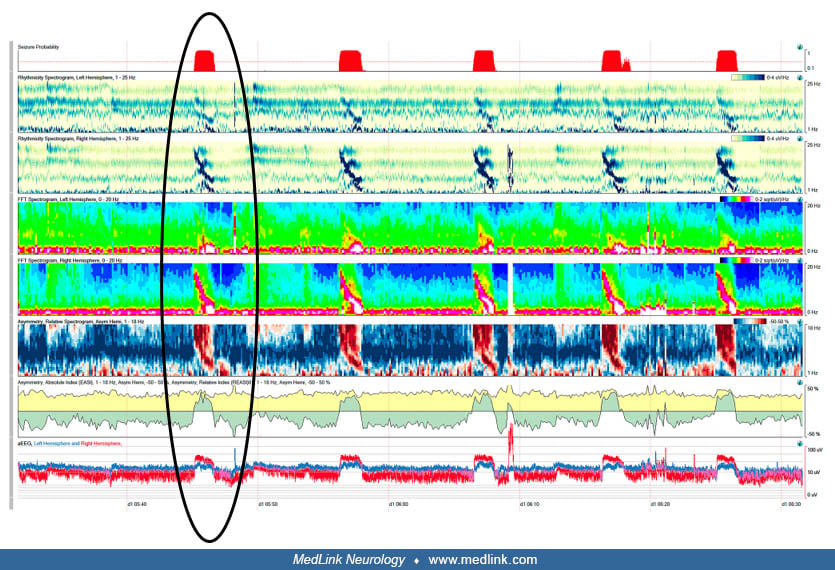
The greatest limitation to EEG in the intensive care unit is the subjective interpretation with suboptimal inter-rater reliability. Quantitative EEG can be used to review large amounts of information generated by cEEG monitoring. Quantitative EEG can analyze long-term trends, allowing a more effective visual analysis that may clarify slowly evolving patterns, such as seizures encountered on cEEG that may be subtle (96).
Quantitative EEG applies the Fourier transformation to identify objective measures of waveform frequency, power, amplitude, asymmetry, and degree of rhythmicity generating numerical values, ratios, or percentages. These are monitored and graphically displayed over long periods of time in a series of trends. Alerting signals may be sent to staff and physicians wirelessly to provide continuous information about the patient’s condition. Their utility in seizure detection is seen regardless of level of training with a sensitivity of at least 80% (87).
There are no anticipated adverse effects when patients undergo a standard scalp EEG. Patients may be uncomfortable during the procedure with the limitations imposed by sustained positioning, provocation techniques, and the time involved performing the study. Rarely, skin irritation from the disc electrodes may be evident following the procedure. This is usually noticeable within the first few days and clears without intervention. Prolonged monitoring may lead to frank skin breakdown at the electrode site. When skin breakdown is severe, ensuring adequate wound care is required, but rarely is a topical antibiotic necessary.
In general, hyperventilation is safe but carries a small risk of inducing potentially adverse effects including seizures or cardiac, respiratory, and cerebrovascular problems. Adverse effects from hyperventilation are expected to be seen in fewer than 3% of patients. Ischemic stroke is a concern in patients with severe cerebrovascular disease and recent stroke (within 3 months) given the risk of hyperventilation-induced vasoconstriction from hypocarbia. Because activating procedures may induce a seizure, patients must provide informed consent prior to performance. In the unlikely event that a seizure occurs, routine seizure management and precautions are instituted.
Invasive electrodes used during intracranial EEG performed as part of a presurgical evaluation carry a risk. In general, for strip electrodes the morbidity is low; infection occurs in 1% of patients. Bleeding is more likely with depth electrodes and stereo-EEG and with larger subdural grids. A greater number of electrodes, longer recording sessions, and more than one cable exit site carry a higher risk that has improved over the years to about 10%. Overall, any complication either immediate or delayed from invasive procedures employing intracranial EEG is low but center-dependent. Therefore, invasive EEG is best handled by a level 4 center (National Association of Epilepsy Centers) with 24-hour neurosurgical support and an on-site intensive care unit in case complications arise.
Special considerations should be observed when performing EEG in certain groups of patients. Pregnant females may require an EEG, and although there is no contraindication to undergoing an EEG, activating procedures should be avoided due to the potential ramifications of an activated seizure on the fetus. Hyperventilation should also be avoided in patients with severe or recent coronary artery and cerebrovascular disease as well as in patients with severe pulmonary disease due to the potential for hyperventilation-induced hypocarbia, leading to vasoconstriction or bronchospasm. Similarly, patients with sickle cell anemia, moyamoya disease, large vessel stenosis supplying the brain, and vasospasm should not undergo hyperventilation procedures given the risk of accentuating ischemia to the point of producing a stroke.
EEG lead placement can change in patients with open traumatic head injuries or craniotomy, and the location should be noted if altered from the 10-20 or 10-10 international system of electrode placement.
Children may have a particularly difficult time tolerating the procedure. Those with intellectual or neurodevelopmental disability may have any combination of language or communication limitations, high levels of anxiety, or behavioral disorders or may manifest sensory hypersensitivities that create barriers and challenges that make it difficult for even the most seasoned technologist to complete a successful EEG. It is essential to provide a soothing and calming influence to comfort a distressed child. Modifications might be made during activating procedures to accommodate the child, including blowing a pinwheel to induce hyperventilation. Photic stimulation is useful in the pediatric population due to potential for provoking generalized spike-and-waves in the form of a photoparoxysmal response that is important not only for diagnosis but also for treatment.
The procedural technique in patients with suspected (or diagnosed) functional seizures (psychogenic nonepileptic attacks) should be a standard scalp EEG recording. Care in documenting any events as typical of the outpatient habitual event is important. Video recording is available on most modern proprietary EEG machines and should be used. Activating procedures should be performed unless otherwise contraindicated as they have proven useful for reproducing nonepileptic events in some patients.
Once nonconvulsive seizures or nonconvulsive status epilepticus have been diagnosed, prolonged EEG monitoring is required. Similarly, generalized or lateralized periodic discharges identified on scalp EEG are special considerations for requiring cEEG. In addition to the prognostic value, the benefits of cEEG include both diagnosis and quantification of nonconvulsive seizures, avoidance of over- and undertreatment of status, and maintenance of suppression-burst on EEG in refractory and super-refractory status.
EEG recording parameters are modified when evaluating brain death. A full set of scalp electrodes are used (except in areas superficial to recent cranial surgery or trauma), and impedance is adjusted to 100 to 10,000 Ohms; the integrity of each electrode is tested, interelectrode distance should be at least 10cm, sensitivity must be increased to at least 2uV/mm, and the test should proceed for at least 30 minutes without blood pressure, temperature, and sedation issues that could interfere with recording (83).
Coupling EEG with the power of computing has been furthered using artificial intelligence. Ever-evolving technological development in artificial intelligence, including machine learning and deep learning methods, is steadily being applied using algorithms that separate the EEG into components to fragment a complex series of waveforms into independent components for analysis and advanced identification. Until recently, algorithms have found only fair to moderate agreement in the interpretation of short segments of abnormal EEG selected for analysis (08). Better results were obtained with comprehensive expert assessment of the entire recording using a large dataset of routine EEG recording. Almost perfect agreement was found for generalized epileptiform abnormalities. In addition, substantial agreement was noted for focal epileptiform discharges, diffuse non-epileptiform abnormalities, and normal EEG records (08). Overall, the algorithm achieved expert-level performance in reading routine clinical EEGs using a convolutional neural network model. Using artificial intelligence can help to avert misinterpretation of EEG, which has been the principal reason for neurologists to render an epilepsy misdiagnosis. In addition, with a rising number of EEG referrals, artificial intelligence can help make the workload more manageable. Importantly, in areas of the world where limited resources exist, artificial intelligence can help address patient access to EEG interpretation without experts available to render a diagnosis.
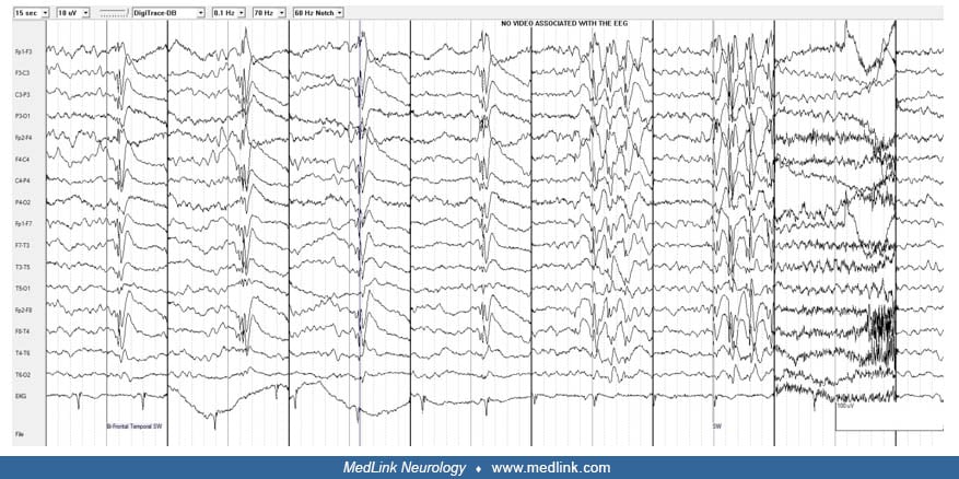
Case history. A 26-year-old man from Kuwait was in good health until he sustained a “grand mal” seizure at 20 years of age. No early risk factors for epilepsy were present. His general and neurologic examinations were normal. An MRI of the brain demonstrated a right temporal lesion consistent with a meningioma. An initial standard EEG was normal, but a repeat sleep-deprived EEG demonstrated intermittent right regional temporal delta slowing. He was treated with phenytoin 400 mg by mouth daily, though adverse effects led to variable compliance and poor control of convulsions. A trial of carbamazepine led to improved compliance, but seizures continued every other month. Lamotrigine was added to carbamazepine, which led to improvement of convulsions though resulted in adverse effects including dizziness and “tics.” He was referred for epilepsy surgery. MRI demonstrated a right middle fossa meningioma. He was subsequently admitted to the epilepsy monitoring unit for video EEG to characterize his seizures. A single seizure was captured following a series of stronger “tics” after partial antiseizure medication reduction. His EEG was initially normal but demonstrated bursts of generalized spike-and-wave discharges overnight.
Conclusion. The interictal EEG demonstrates 4 Hz generalized spike-and-wave and generalized polyspike-and-wave. On video review, there were several myoclonic jerks prior to a generalized tonic-clonic seizure of non-focal origin. He was diagnosed with juvenile myoclonic epilepsy based on his interictal and ictal video EEG monitoring and subsequently underwent a change of his antiseizure medication regimen.
Discussion. This case illustrates several important points. First, it illustrates the importance of the EEG in evaluating patients with drug-resistant epilepsy. Next, it demonstrates the intermittent findings that in this person were two-fold because both focal slowing and generalized spike-and-wave and generalized polyspike-and-wave were present. Additionally, it underscores the importance of the EEG in selecting antiepileptic medication. In this case, both phenytoin and carbamazepine were used resulting in aggravation of his “tics” and ongoing convulsions. Lastly, despite the presence of a structural lesion on brain MRI, which assists in localization and predicting a response to epilepsy surgery, obtaining prolonged EEG with ictal recordings are important in confirming one mechanism for syndromic classification and eligibility for epilepsy surgery. After he was given a definitive diagnosis of juvenile myoclonic epilepsy, he was informed that he was not a candidate for epilepsy surgery. He was followed up by neurosurgery, who recommended conservative management of the lesion. He ultimately became seizure-free after he was transitioned to valproate monotherapy. Serial brain MRIs at 1-year intervals remained stable.
EEG depicts radially oriented dipolar activity derived from different cortical regions. However, two thirds of the cortical convexities lie in the fissures or are in deep regions, resulting in widely distributed topographies. Electrical signals are transmitted down a nerve through depolarization of the neuronal axon. When depolarization occurs at the nerve terminus, a synaptic potential occurs, depolarizing the adjacent nerve dendrite. Excitatory and inhibitory postsynaptic potential are summed and are responsible for EEG waveforms (96). When electroactive volumes are large enough (at least 10 cm2), interictal epileptiform discharges can appear on scalp EEG. Electroactive generators have both positive and negative poles, creating a dipole (63). EEG essentially shows continuously changing voltage fields over different geographic areas across time. Summation of the net orientation of the dipoles within the crown of a cortical gyrus, the area of maximal inflow from the scalp into deeper regions of the brain, shifts from a superficial region to a transverse plane. Mathematical transformation of these currents can produce different 3-dimensional voltage topographies on the scalp (73). Resultant source space where a focal IED originates at the level of the cortical convexity or deep within a sulcus can be differentiated into a net current flow that involves superficial and deeper cortex within a sulcus.
The essential components of an EEG include electrodes, amplifiers, a computer control module, and a display device. Electrocardiogram leads, eye movement monitors, electromyogram, and respiratory monitors are frequently used polygraphic leads that are used in conjunction with EEG commonly during sleep studies. Electrodes are placed on the scalp using the standard international 10-20 system of electrode placement. More detailed coverage of the scalp when performing EEG includes the 10-10 system, which modifies the original 10-20 system and implements a newer alphanumeric nomenclature. More closely spaced scalp electrodes (eg, high-density EEG) have an even higher yield (49). At least 25 electrodes are recommended by the International League Against Epilepsy (ILAE) for a clinical EEG study in adults and children with application of subtemporal electrodes.
The A-P (anterior-posterior) longitudinal bipolar montage is most used as a screening montage and identifies maximal electronegativity based on the presence of “phase reversals,” whereas a reference montage identifies it by the highest amplitude or voltage. A minimum of three montages are utilized during an EEG recording using referential and bipolar recordings according to the American Clinical Neurophysiology Society (79).
The input provided by the EEG technologists is critical at the time of EEG recording. An electrolytic paste is placed between the electrode and the skin and is held in place by adhesive (Collodion), suction, or a cap that conducts the electrical potentials to the amplifiers. Minimal technical requirements for performing EEG have been developed (79). The most common recording setting includes sensitivity (adult = 5 to 15 µV/mm, child = 10 to 20 µV/mm, infant = 15 to 30 µV/mm); standard filter settings of 1 Hz to 70 Hz (notched filter used with 60 Hz artifact) and a time base of 30 mm/second.
Montages follow a common system in which left-sided electrodes are usually depicted higher on the page than right-sided electrodes. The ability to integrate software applications (eg, spike and seizure detection algorithms) and obtain quantitative EEG (compressed spectral arrays) with multimodal trending has extended this capability to the critically ill population (86). Computer-assisted source modeling of dipolar and distributed EEG sources has been helpful for identification of abnormalities in parallel with greater computational ability seen with digital system technology (66).
The use of artificial intelligence, development of software algorithms, and machine learning is exponentially rising in its potential application in EEG. This is especially true for analyzing EEG and is likely to significantly impact the clinical care of people with seizures and epilepsy. As innovations continue at a rapid rate, applications for clinical use are just beginning, including developing and validating an artificial intelligence model with the ability to distinguish abnormal from normal EEG recordings and to classify abnormal EEG recordings into categories relevant for clinical decision-making (108). Deep learning also has potential in automating detection of epileptiform activity and reducing the time and burden of visual analysis that is typically required in clinical practice (20). A logistic regression model trained using spectral and network-derived EEG features using dynamic network models has served to provide initial information to differentiate between epilepsy and non-epilepsy (57). This development could signify an advancement in diagnosing epilepsy even when EEGs do not show interictal epileptiform discharges. Implications include widespread use for clinicians in rural and underserved areas, as well as those interpreting high volumes of EEGs at epilepsy centers, with the use of artificial intelligence in patients with an established diagnosis of epilepsy as well as following a first seizure. Advances in artificial intelligence will impact detection, prediction, and seizure forecasting. This will potentially impact patients with advanced seizure warning systems and those that could guide interventions, changing the role of EEG in clinical practice for patients with epilepsy.
In conclusion, the utility of EEG in clinical practice relies on an accurate interpretation of the recording. Oftentimes, the interpreter is not the treating clinician, and, therefore, optimal reporting is essential to translate the results of the EEG into clinical management of the patient. The moderate interobserver reliability of EEG interpretation may be partly explained by the different reporting styles utilized, and there is significant variability in the observation of guidelines for EEG reporting that couples the science of electrophysiology with clinical practice (07).
All contributors' financial relationships have been reviewed and mitigated to ensure that this and every other article is free from commercial bias.

William O Tatum IV DO FACNS
Dr. Tatum of Mayo Clinic Florida received consulting stipends from Neurelis and consultant fees from Synergy Medical Solutions.
See ProfileSeyed M Mirsattari MD PhD
Dr. Mirsattari of Mayo Clinic has no relevant financial relationships to disclose.
See ProfileBrin Freund MD
Dr. Freund of Mayo Clinic Florida received research contracts or grants from NeuroOne and UniQure and honorariums from Xenon and UCB as a consultant/advisor.
See Profile
John M Stern MD
Dr. Stern, Director of the Epilepsy Clinical Program at the University of California in Los Angeles, received honorariums from Ceribell, Jazz, LivaNova, Neurelis, SK Life Sciences, and UCB Pharma, and Xenon as advisor and/or lecturer.
See ProfileNearly 3,000 illustrations, including video clips of neurologic disorders.
Every article is reviewed by our esteemed Editorial Board for accuracy and currency.
Full spectrum of neurology in 1,200 comprehensive articles.
Listen to MedLink on the go with Audio versions of each article.
MedLink, LLC
3525 Del Mar Heights Rd, Ste 304
San Diego, CA 92130-2122
Toll Free (U.S. + Canada): 800-452-2400
US Number: +1-619-640-4660
Support: service@medlink.com
Editor: editor@medlink.com
ISSN: 2831-9125
Epilepsy & Seizures
Jul. 08, 2026
Neurobehavioral & Cognitive Disorders
Jun. 17, 2026
Epilepsy & Seizures
Jun. 02, 2026
Neuro-Oncology
May. 27, 2026
Movement Disorders
May. 18, 2026
Neuropharmacology & Neurotherapeutics
May. 14, 2026
General Neurology
May. 13, 2026
Neuropharmacology & Neurotherapeutics
May. 11, 2026