


Neurobehavioral & Cognitive Disorders
Mental status examination
Jun. 17, 2026
MedLink, LLC
3525 Del Mar Heights Rd, Ste 304
San Diego, CA 92130-2122
Toll Free (U.S. + Canada): 800-452-2400
US Number: +1-619-640-4660
Support: service@medlink.com
Editor: editor@medlink.com
ISSN: 2831-9125
Toll Free (U.S. + Canada): 800-452-2400
US Number: +1-619-640-4660
Support: service@medlink.com
Editor: editor@medlink.com
ISSN: 2831-9125
Worddefinition
At vero eos et accusamus et iusto odio dignissimos ducimus qui blanditiis praesentium voluptatum deleniti atque corrupti quos dolores et quas.
Hydrocephalus remains a complex clinical entity with treatments ranging from endoscopic third ventriculostomy, shunting (ventricular or lumbar), elimination of the obstruction (such as in the case of mass lesion), or choroid plexectomy (rarely performed alone). When placement of a ventricular shunt is indicated, the distal end of the shunt catheter is most commonly placed in the peritoneum; however, over 36 distal shunt diversion sites have been described in the literature (28). Other common distal catheter placement options thus include the atrium of the heart, pleural space, and gallbladder. Although this is an effective treatment, numerous complications can occur in the operative placement and management of neurosurgical shunts. The latest thoughts on the placement and management of shunts are highlighted in this overview. Additionally, the importance of minimizing radiation exposure in evaluating shunted patients is discussed, including the use of rapid sequence MRI scans for surveillance and emergent evaluation. The potential impact of tablet computers on programmable shunts is also reviewed.
|
• Shunt malfunction is a life-threatening condition and must be treated urgently or emergently. | |
|
• When evaluating for a potential shunt malfunction, it is imperative that the “baseline” image is known to have been taken at a time of shunt function to avoid the misinterpretation of “no change” as the incorrect conclusion of “no evidence of shunt malfunction.” However, one must also use caution as not all patients demonstrate clear ventriculomegaly in the setting of shunt malfunction. | |
|
• Image gently in shunted patients as a lifetime exposure to unnecessary radiation puts them at increased risk of developing leukemia, brain tumors, and other cancers. | |
|
• Attention to detail in the operating room (reduced traffic, preoperative antibiotics, patient preparation, and efficient operating) is key to reducing shunt infections. |
Hydrocephalus, or excessive cerebrospinal fluid, is one of the most commonly encountered neurosurgical conditions.
The first implantable shunt was placed in 1952 by Nulsen and Spitz, and since then, shunt placement has remained the most common treatment of hydrocephalus, despite endoscopic third ventriculostomies with or without choroid plexectomy having made a resurgence in patients (especially in the pediatric population) with obstructive hydrocephalus. (It should be noted that endoscopic third ventriculostomy is typically contraindicated in communicating hydrocephalus.) The peritoneum is the most common location for placement of the distal shunt catheter; however, the distal end is also commonly placed in the right atrium, pleural cavity, or gallbladder. As shunt malfunction can be a life-threatening condition, it is essential to recognize the often progressive and symptomatic signs of shunt failure. This article concentrates on the nature of shunt malfunction, its diagnosis, and management.
Shunt malfunctions. Shunt failures can occur due to obstruction, disconnection, migration, fracture, ascites, abdominal pseudocyst, infections, and even constipation (27). Occlusion of the ventricular catheter is the most common type of mechanical obstruction of shunts and accounts for approximately half of the mechanical obstructions (19). This is typically thought to occur as a result of the choroid plexus growing into the ventricular catheter, although more recent histopathological evidence suggests different types of cellular infiltrates can also cause obstruction, including inflammatory or reactive cells in either early or late shunt failure, respectively (38). It is important to note that the shunt system can occlude anywhere along its course, including the distal catheter, reservoirs, valve, and distal tubing.
Distal obstruction is far less common than proximal obstruction, and the occurrence is more likely to be delayed (14). When distal obstruction is suspected, an abdominal ultrasound study can be useful to look for a pseudocyst, ascites, or constipation. Treatment of infected pseudocysts involves the administration of antibiotics and externalization of the shunt for a brief period. Infection must be ruled out in cases of ascites as CSF ascites can be due to infection, shunt-disseminated metastases, or poor absorption of CSF into the peritoneum (11). Distal obstruction can also occur without an associated infection. In particular, premature infants with a high protein count in their CSF may present with a distal obstruction without associated infection that can often be cleared by irrigating the shunt, obviating the need for an operation.
Disconnection of a shunt rarely occurs early after placement of a new shunt unless there is a technical error. In some shunt systems, the ventricular catheter snaps onto a reservoir, and if this is not done properly, the ventricular catheter can disconnect from the reservoir, resulting in malfunction.
Additionally, the catheter is typically tied with a suture to the valve, but if this is not done, there is a higher risk of disconnection occurring. Late fractures often result from chronic mechanical stress associated with patient growth and movement. Using spectroscopy, conventional histology, and scanning electron microscope, Yamamoto and colleagues found that mineral deposits in degraded shunt tubing consisted of hydroxyapatite (45). The most extensive calcification was seen in the neck, presumably due to the increased mechanical stress in this region. The deposition of sodium chloride and other crystals involving the valve has also been demonstrated as a contributing factor in shunt malfunction (40).
Fractured shunt tubing is typically assessed with a shunt series or x-rays of the head, neck, chest, and abdomen in the anterior-posterior and lateral views. However, fractured catheters can be deceiving on x-ray given the fibrous and calcified tracts that can develop. Thus, if there is clinical concern for shunt malfunction, one must consider surgical exploration. Most surgeons will revise a shunt when a fracture or disconnection is demonstrated because the fibrous tract along a fractured shunt can continue to drain CSF for an indeterminate length of time but will ultimately fail.
The evaluation of a shunt malfunction is, first and foremost, a complete history and physical examination, including a funduscopic examination. Symptomatic complaints of shunt calcification may include pain in the neck or chest wall region, limitation of neck movement due to tethering of the shunt, and cutaneous manifestations such as skin irritation overlying the shunt (37). Unfortunately, children with ventriculoperitoneal shunts are at risk of developing malignancies due to repeated exposure to diagnostic radiation (30). Therefore, the Image Gently campaign has emphasized the importance of reconsidering the need for radiation as well as altering the type of radiation performed, particularly in children (13). Multiple studies support not performing a routine shunt series in every child with a possible shunt malfunction unless clinical evidence suggests a fracture or malposition. One such retrospective longitudinal cohort analysis performed by Antonucci and colleagues, most CT scans obtained in the cohort were not followed by surgical intervention (03). If available, rapid sequence MRI can be used to assess the ventricular size as there is no radiation exposure, and it does not require sedation because it can be performed in less than 1 minute. If rsMRI is not available, consideration of a low-dose CT would be another option for minimizing radiation exposure. In a child with an open fontanelle, ultrasound can also be effective. In any event, careful consideration should be given to ordering diagnostic radiation in these patients. The routine of ordering a shunt series and a CT scan on all cases, often even before notifying the neurosurgeon, must be stopped.
When imaging is employed in the evaluation of shunt malfunction, there may not be a discernable radiographic change in ventricular configuration, particularly if the comparison image is from a previous shunt malfunction. This may then lead to a subsequent misdiagnosis of no current shunt malfunction. Thus, it is imperative that the “baseline” image is known to have been taken at a time of shunt function. However, one must also use caution as not all patients demonstrate clear ventriculomegaly in the setting of shunt malfunction.
As technology advances, new modalities for the evaluation of shunt malfunction become available. One publication describes the use of cerebral regional oxygen saturation monitoring in the assessment of pediatric shunt patients. This technology has most commonly been used for the intraoperative monitoring of children undergoing repair of congenital heart disease. The advantage is that it is a noninvasive, easy-to-apply technology that gives instantaneous readings. The researchers found that in the setting of malfunctioning shunts, there was an asymmetrical hemispheric cerebral regional oxygen saturation, and greater changes in regional oxygen saturation occur for distal versus proximal shunt malfunction (01). Although this methodology requires more investigation, it potentially provides another noninvasive and safe technique for evaluating shunt malfunction.
In shunted adult patients with a diagnosis of normal pressure hydrocephalus, a technique of using noninvasive auditory tests (as currently used for universal neonatal hearing screenings) for the diagnosis of cerebrospinal fluid shunt malfunction was described by Sakka and colleagues (36). Ultrasound-based measurements to characterize the movement of flow through the shunt-valve interface in response to CSF flow is another potential mode of noninvasive testing for shunt malfunction (04).
Surgical principles. Shunt revision usually involves the exploration of a shunt with the replacement of specific malfunctioning components. The use of routine surgical practice guidelines, including extensive skin preparation with an antiseptic to avoid infection, can streamline the entire process, but the treatment of a shunt malfunction is necessarily an individualized matter. Identified risk factors for first-time shunt malfunction include age younger than 6 months at first shunt placement, cardiac comorbidities, and use of an endoscope during proximal catheter placement (35). Additionally, obesity is a growing issue in society today and contributes to some unique complications at the time of ventriculoperitoneal shunt placement or revision. Several papers have been published to address the avoidance of migration of the peritoneal catheter in morbidly obese patients. A technique of placing a stitch beside the peritoneal catheter, which extends through the peritoneum and the posterior and anterior sheath of the rectus abdomens muscle and subcutaneous fat, to eliminate the dead space available for migration has been described by Nakahara and colleagues (29). There have been attempts at generating an increased safety profile for ventriculoatrial shunts for obese individuals as an alternative to ventriculoperitoneal shunts (18).
As above, one of the most common causes of obstruction is choroid plexus (or other tissue) occluding the proximal (ventricular) catheter. Removal of a ventricular catheter from the highly vascularized choroid plexus can result in intraventricular and parenchymal hemorrhages. These hemorrhages increase the risk of subsequent shunt malfunction and can also result in more severe neurologic injury. It is, thus, imperative to coagulate the proximal catheter (bipolar coagulation of the stylet that is inserted into the ventricular catheter) prior to catheter removal; if the catheter cannot be removed, orphaning the proximal catheter may be necessary to reduce risk to the patient.
There is evidence that endoscopic techniques or using a laser filament within the shunt tubing may decrease the need to replace occluded ventricular catheters (17).
One technique for revising the distal catheter was described in which the previously placed distal catheter is exposed at its entry site into the abdomen and a soft guidewire is inserted down the distal catheter into the peritoneum (41). The distal catheter is then removed over the guidewire and a peel-away sheath and dilator are inserted over the guidewire, and the sheath is used to guide the new distal catheter into the peritoneum. It should be noted that the etiology of failure (ie, pseudocyst, abdominal infections, ascites, etc.) may require a new distal shunt location, such as a ventriculoatrial shunt placement.
When revising a shunt, manometry has become the mainstay to test opening ventricular pressure. Furthermore, one should also test fluid “run-off” distally to determine if there is a functioning distal shunt system. It is important to note elevated intra-abdominal pressure as this can also be associated with shunt malfunctions despite a technically “functional” system (15).
Urgent to emergent surgical shunt revision is indicated in any patient with clinical signs of malfunction. It should be emphasized that a shunt malfunction is a potentially life-threatening condition and should be treated as such. Asymptomatic malfunction may be found on routine evaluations either by detecting a fracture in the shunt tubing or by evidence of papilledema on clinical examination. The fibrous tract formed around the tubing can function as a conduit for CSF and, thus, prevent acute symptoms. These tracts are not reliable, and the shunts should be revised in a timely fashion. Asymptomatic individuals with first-time shunt revisions have decreased infection rate and subsequent shunt failure compared to symptomatic individuals with first-time shunt revisions (44).
There are no true contraindications to shunt revision in the setting of shunt malfunction other than an uncorrected bleeding diathesis. Signs of increased intracranial pressure must also be addressed. If there is an active infection, external ventricular drainage may be necessary until the infection is cleared. A suspected infection can sometimes be a relative contraindication to one-stage shunt revision, with relative favoring of externalization or partial removal of the shunt.
The heterogeneity of the disease process complicates outcomes analysis for shunt function. The Shunt Design Trial examined a series of 344 patients prospectively and gave significant data on the value of different valve systems to prevent shunt complications (10). This study had a particular interest in the problem of overdrainage, along with a focused interest on the siphon-resistant and flow-controlled valves on the market. The accumulation of extra-axial fluid as the ventricular system collapses was noted in 3.4% of the patients studied. This may (but not invariably) require surgical treatment by drainage or revision of the valve. A long-term follow-up of data from the Shunt Design Trial by Kestle and colleagues noted that in children younger than 18 years of age, the type of shunt valve did not affect outcomes (20). Additionally, a review of randomized controlled trials examining clinical outcomes for various shunt designs endorsed no significant differences between fixed-parameter and adjustable valves (16). Another overdrainage syndrome of “slit ventricles,” more appropriately called “symptomatic small ventricles,” often presents recurrent problems in patients with ventricles that are small and apparently prone to relapsing shunt malfunctions. Although this is often thought of as a low-pressure type of overdrainage, when these shunts malfunction, the intracranial pressures are frequently high. An important part of the pathophysiological process may be the loss of compliance in the ventricular system, resulting in symptomatic increased intracranial pressure without radiographic change in the size of the ventricle. This can be documented in many patients with the use of mannitol in the inpatient setting. If the symptoms improve along the expected time scale with several repeat boluses of mannitol, treatment of the increased intracranial pressure is necessary.
Different approaches have been proposed to diminish overdrainage, and thereby reinflate the ventricles, allowing treatment of small ventricle symptoms or slit ventricle syndrome. These include the use of siphon prevention or flow control valves; the use of programmable, or adjustable, valves, which can be magnetically reset or “programmed” to have a different flow resistance (43); and subtemporal decompression or another type of cranial expansion. A well-controlled double-blinded study has not been attempted for this complex clinical problem.
Despite being one of the most commonly performed neurosurgical procedures, shunts are consistently associated with high rates of complications requiring subsequent revision. Shunts have previously been estimated to fail at an overall rate of approximately 10% a year, with even higher failure rates of around 30% to 40% within the first year after implantation and up to 50% within the first 2 years after implantation (14). However, the difficulty in calculating a precise estimate of overall failure rates lies in the varied definitions of shunt failure throughout the literature, as well as variable time to follow-up and study duration. A 2023 meta-analysis of 46 studies dealt with these concerns by ultimately considering shunt failure as equivalent to shunt revision and then utilizing survival analysis of shunts that remain revision- or failure-free within 6 months. The resultant pooled prevalence of overall shunt failure in adults was 22.7% (32). Notably, this study did not investigate pediatric outcomes, although multiple studies have previously established significantly greater failure rates in children, particularly in patients younger than 1 year old. One such study collected data on 773 operations and found a substantially higher failure rate for shunts inserted in the first 6 months of life (35% to 47%) compared with shunts placed in children older than 6 months of age (14%) (09). Similarly, a single-center study that reviewed 727 operations over a 13-year period also found a higher failure rate associated with young children (31). Additionally, this study found higher failure rates in cases involving complex shunts (with multiple proximal catheters) compared to simple “linear” shunts, as well as when there was a short interval between revisions.
One multicenter evaluation of over 5000 operations over a 2-year period found that 35.7% of shunt failures were potentially preventable, with the most common etiologies being infection (44%) and proximal catheter malposition (27%) (08). Predictors of preventable shunt failure also include lack of endoscopy, recent shunt infection, shunt type, and center (08). Other factors contributing to shunt failure or malfunction include the etiology of hydrocephalus, type of hydrocephalus, and previous treatments before shunt surgery (34; 02).
In children undergoing a shunt revision, cardiac risk factors, a chronic history of seizures, and a history of neuromuscular disease were independently associated with shunt failure (12). In a retrospective study of 74,552 pediatric patients with a CSF shunt, peritonitis, papilledema, and oculomotor palsies were the strongest indicators of shunt malfunction requiring a visit to the emergency department (33). Finally, one study of a pediatric population also indicated that the number of surgeons in the operating room was associated with shunt failure.
The most common and serious risk of shunt surgery is shunt infection, which occurs in the operating suite from airborne bacteria, the patient’s skin flora, or a break in sterile technique. Kestle and colleagues published a “shunt infection protocol” in which they demonstrated a reduction in the rate of postoperative shunt infections from 9% to 6% after implementation of such protocol, including (21):
• sign on the door to restrict traffic | |
• head turned away from operating room door | |
• preoperative antibiotics (cefazolin or vancomycin) | |
• hair clipping and local site cleaning | |
• hand washing with betadine or chlorhexidine | |
• surgeon double-gloving | |
• ioban drape placed on the patient | |
• injection of gentamycin or vancomycin into the reservoir | |
• a single postoperative dose of antibiotics. |
There is growing evidence that supports the use of an antibiotic-impregnated shunt to reduce the infection risk. In a study of 1605 patients in the United Kingdom, antibiotic shunts resulted in a lower rate of infection and reduced cost compared to the use of a standard shunt (25; 26). It is worth noting that the study found no difference in shunt failure rate between an antibiotic shunt and a standard shunt.
Intracranial hemorrhage during surgery is an uncommon but serious complication. Savitz and colleagues noted an approximate 4% rate of intraparenchymal or intraventricular hemorrhage in the absence of coagulopathy (39). As above, this typically occurs when the choroid plexus invades the ventricular catheter and results in bleeding when the ventricular catheter is removed. Other risks associated with shunt insertion to be considered and discussed with patients and families are seizures, malpositioning of either the proximal or distal shunt catheters, and skin breakdown over the hardware.
Hydrocephalic women who become pregnant could develop symptoms of increased pressure due to diminished intra-abdominal reserve in resorbing fluid and increased intra-abdominal pressure. Fortunately, this is an uncommon problem. Diagnostic imaging in this case would favor the use of MRI over CT (to avoid fetal radiation), but surgical management in the case of true malfunction would be as in the nonpregnant patient. Endoscopic third ventriculostomy may be effective in this setting.
Patient 1. A 7-year-old girl with a history of hydrocephalus secondary to a perinatal intraventricular hemorrhage presented with a headache that progressed over 24 hours, together with nausea and vomiting. There was no papilledema, but a CT scan showed ventricular distention when compared with an earlier baseline symptom-free scan.
At surgery, a proximal shunt obstruction was identified, and after replacement with a new ventricular catheter, shunt function normalized, and her symptoms resolved.
Patient 2. A 3-month-old infant with severe hydrocephalus from congenital aqueductal stenosis presented with irritability and fever; CT scan was performed.
Collapse of the ventricular system from overdrainage was noted, with bleeding into the subdural space. Overdrainage was treated without surgery by keeping the patient flat to avoid a gravitational siphoning effect. The subdural blood, a sign of overdrainage, resolved.
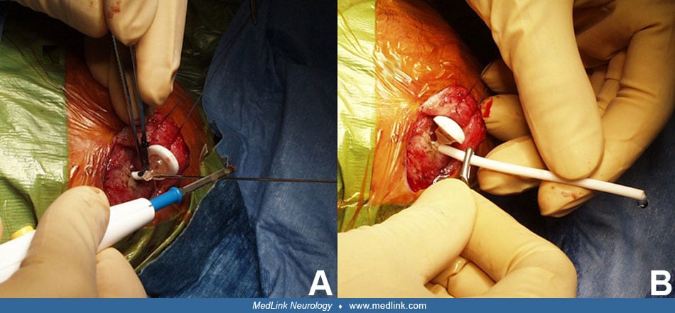
The structure of a ventriculoperitoneal shunt. Virtually all shunts for hydrocephalus are made from medical-grade silastic, and all but a few (used in small infants or special circumstances) contain an in-line valve.
There are many brands and varieties of shunt equipment, and although surgeons tend to have brand loyalty based on experience, none has proven to be superior to any other brand. The one exception might be the programmable valve, which can be particularly useful in the setting of normal-pressure hydrocephalus in adults. In these patients, the pressure of the valve can be manipulated without surgical intervention by simply placing a programming wand over the valve. A meta-analysis conducted by Li and colleagues demonstrated that the use of programmable valves is not only safe but may actually reduce the revision rate and complications of under- or overdrainage, particularly in individuals less than 18 years of age with first-time shunt placement (23). Of note, tablet computers have been shown to unintentionally change the setting of programmable shunts if held near the valve, and patients need to be educated about this possibility (42).
Most valves act as simple resistors and rectifiers to flow. This means that flow in the reverse direction is blocked by a check valve, and the presence of flow depends on sufficient elevation of pressure above the valve in the lumen of the shunt. Therefore, the absolute pressure within the intracranial end of the shunt system may depend significantly on body position as the drainage, or “siphoning,” of fluid in the distal part of the tubing is greatest in the upright position. This siphoning effect can be useful in premature infants with soft malleable skulls. Siphoning was recognized as a potential cause of symptoms years ago; thus, the “antisiphon” device was created. This device makes the flow dependent on the absolute difference in pressure between the upper chamber and the lower chamber, and it also either closes the valve or significantly increases the resistance to flow, if actual downstream pressure becomes subatmospheric. Correct functioning of this part of the device depends on the mobility of a membrane within it, along with access of the diaphragm to the external air pressure. Thus, such devices may fail under a thick scalp. If they become trapped in the closed position, they inhibit flow and create a situation otherwise identical to a shunt malfunction. Liniger and colleagues found no difference in the development of slit ventricles or slit ventricle syndrome with flow control versus antisiphon valves (24).
Shunt failure and CSF physiology. The majority of symptoms related to untreated hydrocephalus are a result of increased pressure. Thus, the symptoms and signs are similar to those of hydrocephalus itself: headache, nausea, vomiting, upgaze paralysis, and papilledema. In a prospective study, drowsiness was found to be the best predictive symptom (05). In infants, a full fontanelle and head growth crossing percentiles can also be observed. Usually, with higher pressure, the ventricles get larger, but the quantitative degree varies significantly from patient to patient. However, in the patient with a poorly compliant brain, ventricles may not get larger at the time of a shunt malfunction. Although this situation is not common, it usually occurs in patients with “slit ventricles” or shunted arachnoid cysts. Additionally, up to 15% of pediatric patients have altered brain compliance and may not display enlargement of their ventricles in the presence of shunt failure and increased intracranial pressure (14). In some cases, shunt malfunction is difficult to diagnosis both clinically and by CT scan. Shuntograms may be useful in this regard. There are no validated prediction models for shunt malfunction in low-risk individuals based on clinical presentation alone (07).
There has been much discussion about “shunt” allergies. If this occurs, it is rare. The typical presentation of an allergy to silicone shunts includes abdominal pain and irritation or breakdown of the skin overlying the shunt. It is typically diagnosed via the detection of antisilicone antibodies and can be treated by replacement of the silicone shunt with a polyurethane shunt (22). CSF eosinophilia is also a useful marker in determining shunt malfunction secondary to a hypersensitivity reaction or bacterial infection.
Loculation. Because the ventricular system can be hydrodynamically divided by septa into several compartments, it occasionally happens that an isolated part of the ventricular system enlarges despite adequate shunting of a portion that does not communicate with this. This usually occurs in the setting of infection. Such shunt failure cases may require the placement of more than one catheter or fenestration of the obstructing tissue endoscopically to make the system unified. The latter procedure permits drainage through a single catheter, which is often preferable. Endoscopic third ventriculostomies have proven to be efficacious for the management of childhood hydrocephalus due to congenital or acquired aqueductal stenosis but are less effective in the management of hydrocephalus secondary to inflammatory or infectious etiologies (06).
All contributors' financial relationships have been reviewed and mitigated to ensure that this and every other article is free from commercial bias.
Michael J Schneck MD
Dr. Schneck of Loyola University of Chicago has no relevant financial relationships to disclose.
See ProfileAlexandria Pecoraro MD
Dr. Pecoraro of Loyola University Medical Center has no relevant financial relationships to disclose.
See Profile
Matthew Lorincz MD PhD
Dr. Lorincz of the University of Michigan has no relevant financial relationships to disclose.
See ProfileNearly 3,000 illustrations, including video clips of neurologic disorders.
Every article is reviewed by our esteemed Editorial Board for accuracy and currency.
Full spectrum of neurology in 1,200 comprehensive articles.
Listen to MedLink on the go with Audio versions of each article.
MedLink, LLC
3525 Del Mar Heights Rd, Ste 304
San Diego, CA 92130-2122
Toll Free (U.S. + Canada): 800-452-2400
US Number: +1-619-640-4660
Support: service@medlink.com
Editor: editor@medlink.com
ISSN: 2831-9125
Neurobehavioral & Cognitive Disorders
Jun. 17, 2026
Neuro-Oncology
May. 27, 2026
Neuropharmacology & Neurotherapeutics
May. 14, 2026
Neuro-Oncology
Apr. 30, 2026
Neuropharmacology & Neurotherapeutics
Apr. 23, 2026
Neuropharmacology & Neurotherapeutics
Apr. 20, 2026
Neuro-Ophthalmology & Neuro-Otology
Apr. 07, 2026
General Neurology
Apr. 06, 2026