Neurobehavioral & Cognitive Disorders
Mental status examination
Jun. 17, 2026
MedLink, LLC
3525 Del Mar Heights Rd, Ste 304
San Diego, CA 92130-2122
Toll Free (U.S. + Canada): 800-452-2400
US Number: +1-619-640-4660
Support: service@medlink.com
Editor: editor@medlink.com
ISSN: 2831-9125
Toll Free (U.S. + Canada): 800-452-2400
US Number: +1-619-640-4660
Support: service@medlink.com
Editor: editor@medlink.com
ISSN: 2831-9125
Worddefinition
At vero eos et accusamus et iusto odio dignissimos ducimus qui blanditiis praesentium voluptatum deleniti atque corrupti quos dolores et quas.
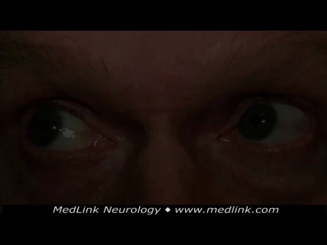
Nystagmus is the term used to describe involuntary rhythmic oscillations initiated by a slow drift of the eyes. Such oscillations can be physiologic; they can usually be differentiated from pathologic nystagmus by the oscillatory features alone. Pathologic nystagmus is usually a manifestation of brainstem or cerebellar dysfunction, although impaired vision may be a contributing factor, especially with onset early in life. Impairments in neuromuscular transmission and ocular motor palsies may also rarely produce pathologic nystagmus. Localization of the underlying disorder depends on identifying the pattern of the oscillations in conjunction with the setting and accompanying neurologic manifestations. This article reviews the common forms of nystagmus, including their clinical features, mechanisms, mimickers, evaluation, and treatment.
|
• Nystagmus refers to rhythmic involuntary oscillations of the eyes. | |
|
• Nystagmus may be physiologic or pathologic, congenital or acquired, pendular or jerk. | |
|
• It should be differentiated from other types of ocular oscillations, including saccadic intrusions and other “nystagmoid” movements. | |
|
• Identifying the pattern of the oscillations is critical to diagnosis. | |
|
• Pharmacologic agents and other interventions are sometimes effective in dampening the nystagmus and lessening associated visual symptoms. |
The word “nystagmus” originates from the Greek word nustagmos, meaning “drowsiness” or “nodding.” The oscillations of the eyes in nystagmus were likened to the nodding head movements of sleepiness (31).
Nystagmus is described by the oscillatory waveform and the plane of its trajectory, the oscillatory amplitude and frequency, the gaze position in which the oscillations occur, whether the eyes move in the same direction (“conjugate”) or opposite directions (“disconjugate”), and whether the oscillations involve one eye or both. The examiner must determine whether the oscillations represent nystagmus or an imitator, whether the nystagmus is physiologic or pathologic, and whether the oscillations are spontaneous or induced by a provocative maneuver.
Waveform. The nystagmus waveform may pendular or jerk. In pendular nystagmus, the waveform consists of two slow phases. In jerk nystagmus, the initial movement of the eyes is slow, but the recovery movement is fast. By convention, the direction of jerk nystagmus is based on the direction of the recovery fast phase. For example, if the fast phase is leftward, the patient has a “left-beating nystagmus.”
Plane of trajectory. The trajectory of the oscillations may be in the horizontal, vertical, or torsional plane. In many cases of jerk nystagmus, the eyes will move simultaneously in two or more planes. In some cases of pendular nystagmus, the trajectory is complex, such that the eyes move in a circular or oval (elliptical) pattern.
Amplitude and frequency. The amplitude of the oscillations refers to the width of the ocular swings; the frequency refers to the speed of the oscillations. Amplitude and frequency are inversely related. When amplitude is low, frequency is apt to be high; when amplitude is high, frequency is usually low. The amplitude and frequency are usually equal in the two eyes, but in some cases, they are markedly different (“dissociated nystagmus”).
In nystagmus with onset after childhood, a high amplitude is often accompanied by the sensation that viewed objects are moving (“oscillopsia”). In adult-onset jerk nystagmus, the oscillopsia consists of a unidirectional movement of viewed objects in the direction of the fast phase. In adult-onset pendular nystagmus, objects will appear to be moving back and forth in the plane of the oscillation. Congenital (infantile) nystagmus gives rise to blurred vision but never to oscillopsia, perhaps because early onset nystagmus leads to a sensory adaptation. Eyes with low-amplitude, high-frequency nystagmus will look like they are shaking or shimmering. In such cases, it is especially challenging to distinguish nystagmus from saccadic intrusions.
In some cases, the nystagmus amplitude will differ markedly between the eyes. An example is the acquired pendular nystagmus of multiple sclerosis, in which the amplitude will be greater in the eye with the more profound optic neuropathy. In spasmus nutans, the amplitude in one eye may be so low that the nystagmus is assumed to be monocular.
Gaze position. Nystagmus may occur in any gaze position. In this regard, two observations are critical to diagnosis: the gaze position in which nystagmus occurs and whether the nystagmus features vary with changing gaze position. For example, congenital (infantile) nystagmus has a horizontal trajectory in all fields of gaze (“uniplanar”), whereas most acquired gaze-evoked jerk nystagmus associated with brainstem or cerebellar disease has a horizontal-rotary trajectory in side gaze and an upbeat nystagmus in upgaze (“multiplanar”). In many forms of acute peripheral vestibular nystagmus, the jerk nystagmus is in the same direction regardless of whether the eyes are in right gaze or left gaze (“unidirectional”). By comparison, in the nystagmus of brainstem or cerebellar lesions, the fast phase of gaze-evoked jerk nystagmus switches directions as the gaze shifts from side to side (“direction-changing”).
Conjugate or disconjugate? In most forms of nystagmus, the two eyes are moving in the same direction (“conjugate”). However, seesaw nystagmus is an example of “disconjugate” nystagmus; one eye rises and intorts while the other eye falls and extorts. Oculomasticatory myorhythmia, a form of nystagmus associated with Whipple disease, displays cycles of ocular convergence and divergence. In the ocular convergence-retraction clonic movements associated with pretectal or dorsal midbrain dysfunction, the eyes converge and retract into the orbit.
One eye or both eyes? Most nystagmus involves both eyes, but there is a notable exception: the monocular (usually vertical) pendular nystagmus of childhood associated with an ipsilateral optic neuropathy usually caused by a diencephalic astrocytoma. Superior oblique myokymia, an imitator of nystagmus, consists of arrhythmic intorsion movements limited to one eye.
Physiologic or pathologic? Physiologic nystagmus occurs in normal subjects, sometimes evoked or exacerbated by fatigue. It can usually be separated from pathologic nystagmus on the basis of its oscillatory features.
Spontaneous or induced? Spontaneous nystagmus occurs without provocative maneuvers; induced nystagmus is provoked by maneuvers useful in diagnosis.
Physiologic gaze-evoked nystagmus has five oscillatory features that, if taken together, usually distinguish it from pathologic gaze-evoked nystagmus: (1) horizontal jerk pattern, often with a slight torsional component; (2) limited to far extremes of horizontal gaze to both sides; (3) low and symmetrical amplitude in the two eyes, although the abducting eye may show slightly greater amplitude (07; 25; 20); (4) similar amplitude in right and left gaze; (5) no more than four beats (“unsustained”).
Because these features may sometimes occur in mild forms of pathologic gaze-evoked nystagmus, the examiner should always exclude the presence of other pertinent manifestations, particularly ocular misalignment, saccadic pursuit, ataxia, and the use of anticonvulsants and sedatives. Pendular nystagmus or jerk nystagmus in the vertical or purely torsional plane should never be considered physiologic.
Caloric nystagmus. This form of jerk nystagmus is induced by the instillation of water into the external auditory canal. With the patient’s head elevated 30 degrees above the horizontal plane, water is instilled into the ear via a syringe without a hypodermic needle. This maneuver creates an ipsilateral current in the endolymph of the horizontal semicircular canal and asymmetric activation of the horizontal vestibulo-ocular reflex on the two sides of the brainstem. When warm water is instilled into the left ear, the left vestibular system is rendered more active than the right. As a result, the eyes drift into right gaze, followed by fast recovery movements toward left gaze (called “quick phases” in this setting). This ocular oscillation is a “left-beating” nystagmus. In clinical practice, the temperature of the instilled water is cold--chilled by ice. It ablates ipsilateral vestibular signaling so that the eyes drift toward the irrigated ear, followed by recovery quick phases that are automatically generated in the opposite direction, creating a jerk nystagmus beating away from the irrigated ear. The COWS mnemonic describes the direction of the nystagmus fast phase after cold water irrigation: “cold opposite, warm same.”
Caloric testing is used primarily in the determination of brainstem death. In comatose patients with intact brainstem circuitry, cold water instillation induces an ipsilateral slow phase conjugate drift of the eyes without recovery fast phases. If the brainstem is not functioning, there will be no observable eye movements.
The caloric irrigation test can also be used in a patient with depressed consciousness to determine if a dilated pupil is a postganglionic manifestation (eye trauma, exposure to topical parasympatholytic agents) or part of an ipsilateral third nerve palsy. In patients with an extra-axial third nerve palsy but a functioning brainstem, instillation of cold water in the contralateral ear will induce full abduction of the eye on the side of irrigation but reduced or absent adduction of the eye on the opposite side. Inasmuch as caloric irrigation functions by stimulating the vestibulo-ocular reflex, why not perform the simpler oculocephalic maneuver in these settings? Because the oculocephalic maneuver provides a much weaker vestibulo-ocular reflex stimulus than does caloric irrigation.
Optokinetic nystagmus. The optokinetic reflex is elicited by moving a striped stimulus across the patient’s visual field. A tape or drum consisting of alternating white and black or red stripes (now also available as a smartphone application) is placed at a distance of 3 meters from the patient’s eyes and moved in the horizontal or vertical plane. In an alert and fixating patient with at least finger-counting visual acuity, the moving stripes will elicit repetitive ipsilateral conjugate pursuit movements that are followed by recovery quick phases. This response is virtually automatic, suppressible only by a strong “defocusing” effort. The great utility of this test is that it elicits repetitive saccades that can be observed for amplitude. A pathologic response is the absence of nystagmus or a reduced amplitude.
The optokinetic nystagmus test is used in five settings: (1) eliciting a normal response in a patient suspected of factitious binocular vision loss; (2) eliciting reduced nystagmus amplitude in the adducting eye in subtle internuclear ophthalmoplegia when the stripes are moved horizontally (25); (3) eliciting reduced nystagmus in the abducting eye in subtle sixth nerve palsy when the stripes are moved horizontally; (4) evoking convergence retraction in dorsal midbrain (pretectal) dysfunction when the stripes are moved in the downward direction; (5) eliciting relatively reduced horizontal nystagmus amplitude when the stripes are moved in the direction of a parietal lesion (40).
Other forms of induced nystagmus. Nystagmus can also be induced by applying high-plus (Frenzel) spectacles, head-positioning, headshaking, mastoid vibration, external auditory canal insufflation, hyperventilation, and loud noises. These maneuvers are designed to draw out various types of peripheral vestibular nystagmus.
Pathologic nystagmus is usually an expression of brainstem or cerebellar dysfunction, but it may also be caused by disorders of vision or peripheral ocular motor dysfunction. Five mechanisms are invoked to explain nystagmus: (1) vestibular tone imbalance created by a left-right imbalance in peripheral inputs to the vestibulo-ocular reflex; it underlies acute peripheral vestibular nystagmus; (2) impaired eccentric gaze-holding by the brainstem neural integrator responsible for the “step” innervation of the extraocular muscles; it underlies gaze-evoked nystagmus; (3) impaired vision, which interferes with stable fixation; it contributes to congenital sensory nystagmus, monocular pendular nystagmus of childhood, and probably to the acquired pendular nystagmus of multiple sclerosis; (4) overshoot of the abducting eye contralateral to weak adduction in internuclear ophthalmoplegia; (5) weakness of extraocular function or fatiguability of neuromuscular transmission; it underlies the nystagmus of ocular motor palsy and myasthenia gravis. The mechanism underlying the many remaining types of nystagmus is unsettled.
For classification purposes, it is useful to divide pathologic nystagmus into pendular and jerk forms and then further divide them into those with onset in early childhood and those with onset later in life.
Early childhood-onset pendular nystagmus.
Monocular. This form of nystagmus has a prominent vertical trajectory (“Heimann-Bielschowsky” phenomenon). It is associated with an ipsilateral optic neuropathy, usually caused by a diencephalic pilocytic astrocytoma (09). Ocular causes of severe monocular vision loss, including amblyopia, may also underlie this form of nystagmus.
Multivector. This form of nystagmus is binocular and often has a circular or elliptical track because there are horizontal and vertical vectors (“multivector”). It is usually a manifestation of a hypothalamic astrocytoma that may produce a lack of appropriate weight gain to the point of inanition (“Russell diencephalic syndrome”) (36). Other causes of this type of childhood multivector nystagmus are the leukodystrophies of Pelizaeus-Merzbacher disease (35) and peroxisomal disorders (20).
Seesaw. This nystagmus is unusual in being disconjugate: one eye rotates upward and outward while the other eye rotates downward and inward.
Often accompanied by bitemporal hemianopia, this form of nystagmus may be a sign of a parasellar lesion (astrocytoma, craniopharyngioma) that infiltrates or compresses the optic chiasm. Alternative causes are septo-optic dysplasia, achiasma, and other forebrain dysgeneses. The mechanism of this nystagmus may be based on a disruption of the anterior visual pathway and mesodiencephalic circuitry (23).
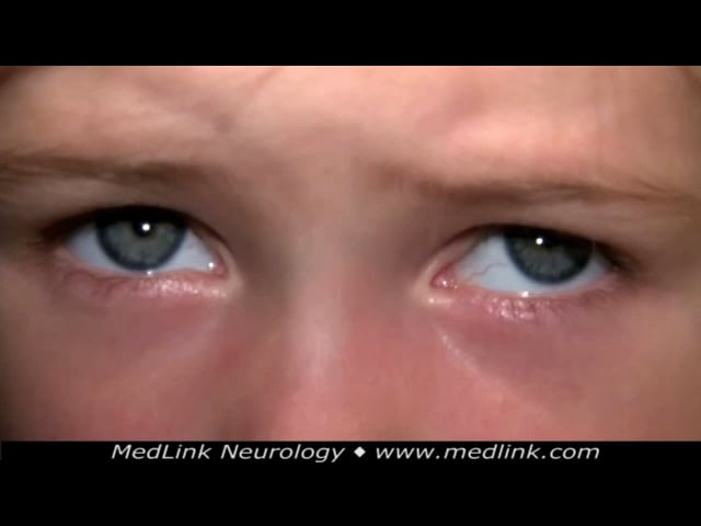
Spasmus nutans. This nystagmus is intermittent with a trajectory that varies from horizontal to vertical. At moments, the two eyes move disconjugately. Amplitude is so low and frequency so high that the eyes will appear to be “shimmering.” The oscillations are usually binocular, but sometimes the motion of one eye is so small that it may be overlooked.
The term “spasmus” relates to the intermittency of the rapid nystagmus, and the term “nutans” refers to the accompanying head and truncal titubation, which is a prominent feature. A third feature is torticollis.
Spasmus nutans has its onset usually between 4 and 14 months of age with spontaneous resolution by age 3 years, leaving no traces. Full neurologic evaluation has never turned up any abnormalities, but such evaluation is necessary because this nystagmus is difficult to distinguish from other forms of pendular nystagmus (28; 02) and opsoclonus (01).
Later-onset pendular nystagmus.
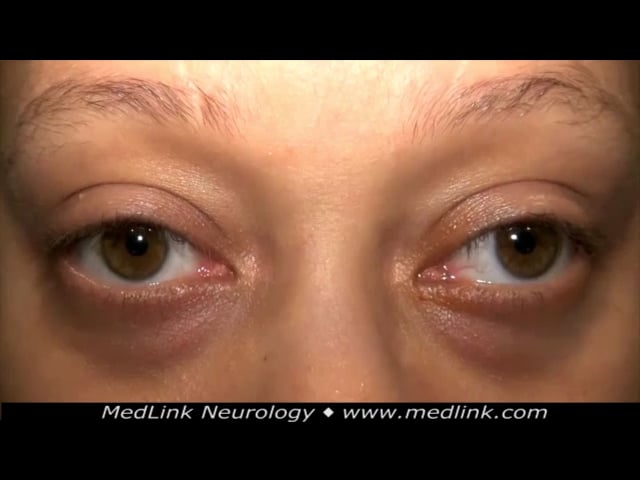
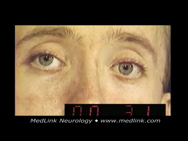
Multivector. This nystagmus, which has a circular or elliptical trajectory owing to horizontal and vertical vectors, looks like the nystagmus of childhood diencephalic disease, but it is recognized later in life in conjunction with chronic multiple sclerosis manifesting brainstem and optic nerve dysfunction. The amplitude of the nystagmus is generally higher in the eye with lesser vision.
Oculopalatal tremor. This nystagmus resembles that of multiple sclerosis except that it often has a more prominent vertical component. It follows by several months a hemorrhage or ischemic stroke affecting the Mollaret triangle (dentate nucleus, inferior olive, and red nucleus). The accompanying features provide the diagnosis: the palate and other branchial arch-derived muscles, including the platysma, trachea, and diaphragm, oscillate synchronously with the eyes.
In addition to displaying the original lesion in the Mollaret triangle, MRI typically also shows new enlargement and high FLAIR/T2 signal in the inferior olive.
Convergent. The eyes converge rhythmically and synchronously with clenching of the jaw muscles (“oculomasticatory myorhythmia”) (29). It is considered pathognomonic of Whipple disease affecting the mesodiencephalon.
Early-onset jerk nystagmus.
Congenital. The most common type of early-onset jerk nystagmus is called “congenital nystagmus,” or “infantile nystagmus syndrome” because it may not be detected until several months after birth (27). In primary gaze position, the waveform may appear to be horizontal pendular, but eye movement recordings show it to be an increasing velocity jerk nystagmus. On side gaze, the oscillation converts to a jerk nystagmus beating in the direction of gaze (“direction-changing”). The nystagmus trajectory remains horizontal in upgaze (“uniplanar”). When nystagmus is of indeterminate duration, this uniplanar feature alone is enough to distinguish it from the later-onset nystagmus of brainstem or cerebellar origin, which typically beats upward in upgaze (“multiplanar”).
Nearly all patients with congenital nystagmus have subnormal visual acuity in both eyes, ranging from 20/40 to 20/400. Because of this impaired vision, they will eventually report blurred vision, but never oscillopsia, another important feature that distinguishes this form of nystagmus from acquired types. Visual function does not decline over time unless a new cause develops. Head-nodding (“titubation”), present in many cases, may falsely suggest a pervasive neurologic disturbance.
Visual acuity is often optimal in a narrow zone of eccentric horizontal gaze on one side, where the eyes enter a “null zone” of reduced nystagmus amplitude. When such a null zone is present, and if visual acuity improves when the eyes are placed within it, the patient may adopt a face turn for straight-ahead viewing. These phenomena justify an extraocular muscle surgery that repositions the eyes to allow a normal head position with improved acuity.
Another feature of this form of nystagmus is that the oscillatory amplitude often markedly reduces with convergence, which may account for improved acuity for viewing near targets. This phenomenon is the basis for another extraocular muscle procedure that may improve visual acuity.
Congenital nystagmus is often idiopathic (“congenital motor nystagmus”), although retinal foveal irregularities have been discovered on optical coherence tomography. However, in at least half of cases, there are underlying ocular abnormalities, such as photoreceptor dystrophy, albinism, aniridia, amblyopia, high refractive error, media opacities, and optic nerve hypoplasia (“congenital sensory nystagmus”) (20). Discovery of these underlying sensory abnormalities may not lead to effective treatment, but they should be identified for prognostic reasons alone and as a basis for genetic counseling.
The features of congenital nystagmus remain stable over a lifetime. Visual aids are helpful. As this condition is unassociated with intracranial abnormalities, brain imaging is not necessary unless it is accompanied by an afferent pupil defect, optic disc abnormalities, or other neurologic manifestations (10).
A common accompaniment of congenital nystagmus is called “latent nystagmus” or “fusion maldevelopment nystagmus.” It is a type of congenital jerk nystagmus present only when one eye is viewing. Covering the other eye produces a conjugate jerk nystagmus directed away from the occluded eye. This phenomenon may be isolated, but more often it is associated with congenital nystagmus and congenital esotropia. A benefit of identifying latent nystagmus is that it labels the accompanying nystagmus (and esotropia) as congenital.
Latent nystagmus is often first recognized only during the first formal testing of visual acuity, when the normal procedure of covering the untested eye brings about oscillation in the tested eye that degrades its visual acuity. To avoid having latent nystagmus interfere with visual acuity testing, the examiner should not occlude the untested eye but, rather, “fog” its acuity with a high-plus lens, which will not elicit nystagmus in either eye.
A rare variant of latent nystagmus has the patently contradictory term “manifest latent nystagmus.” In that variant, monocular vision causes the nystagmus to be present even without occluding an eye (21).
Periodic alternating. Periodic alternating nystagmus, which may be detected early in childhood or later in life, consists of a horizontal jerk oscillation whose direction shifts periodically. After 90 seconds, the nystagmus amplitude gradually lessens until the eyes are still for about 15 seconds. The nystagmus amplitude then gradually ramps up in the opposite direction for 90 seconds with gradually reducing amplitude until the eyes are still again.
The mechanism for this intriguing cyclical nystagmus is uncertain. When periodic alternating nystagmus has its onset in early childhood, it is probably a variant of congenital nystagmus, with similar causes.
Later-onset jerk nystagmus.
Gaze-evoked horizontal (“side beat”). Pathologic horizontal gaze-evoked nystagmus is the most common form of acquired nystagmus. It consists of a horizontal jerk nystagmus limited to eccentric gaze to both sides, the eyes beating in the direction of gaze (“direction-changing”).
This form of nystagmus can be distinguished from physiologic nystagmus because it is sustained, of greater amplitude, and present at lesser degrees of eccentric gaze. It reflects impairment of the brainstem integrator responsible for maintaining eccentric horizontal gaze. When the integrator fails, elastic orbital forces drive the eyes back toward the primary position. That inward movement is met by compensatory fast-phase eye movements attempting to return the eyes to the desired eccentric gaze position (20). Etiologies include toxic-metabolic processes, including sedatives and anticonvulsant medications, and lesions in the brainstem or cerebellum.
Many patients with horizontal gaze-evoked nystagmus also have an upbeat nystagmus in upgaze. The combination of this upbeat nystagmus and direction-changing on side gaze distinguishes this form of nystagmus from peripheral vestibular nystagmus, which always beats in the same direction regardless of gaze position.
An uncommon feature of gaze-evoked nystagmus is “rebound nystagmus.” When the eyes return to the primary position after prolonged eccentric gaze, they display a brief conjugate jerk nystagmus in the opposite direction. This phenomenon localizes the lesion to the medulla, but it can be mimicked by myasthenia gravis (20).
Downbeat. Downbeat nystagmus refers to a conjugate jerk nystagmus with the fast phase beating downward. It is easiest to appreciate when the eyes are in down-and-lateral gaze (38). Because a slight horizontal trajectory is combined with the more dominant downward trajectory, the resultant vector will be an oblique downward movement (“side pocket nystagmus”).
The underlying mechanism in downbeat nystagmus may be impaired downward pursuit owing to dysfunction of the cerebellar floccular and parafloccular lobes (15; 37; 38). Among structural abnormalities evident on brain imaging, the most common are hindbrain tumors, strokes, and dysplasias, including Chiari malformation (06). Nonstructural etiologies include hereditary, alcoholic, and autoimmune (including paraneoplastic) spinocerebellar degenerations, anticonvulsant and lithium medication use (05), brainstem encephalitis (13), Wernicke encephalopathy, and magnesium and vitamin E deficiency (11; 39). Downbeat nystagmus may be unassociated with other neurologic manifestations and remain idiopathic, although ataxia often appears later as part of advancing cerebellar degeneration (11; 37).
Upbeat. This vertical jerk nystagmus present in primary gaze typically increases in amplitude in extreme upgaze.
It is a sign of acquired disease anywhere from the medulla to the midbrain. Wernicke encephalopathy is an important cause, as is multiple sclerosis. Degenerative conditions, tumors, strokes, intoxications, and inflammatory processes may also be responsible. When nystagmus amplitude atypically increases in downgaze, the lesion lies in the lower brainstem (08). This form of nystagmus should be distinguished from the more common form of upbeat nystagmus that is not present in the primary gaze position and is evoked only by upgaze. It accompanies gaze-evoked nystagmus and may also be seen in dorsal midbrain syndrome and myasthenia gravis.
Pure torsional (rotary). In this type of jerk nystagmus, the trajectory is purely torsional (rotary).
It derives from lesions of the brainstem or cerebellum, most commonly the medulla (26). In some cases, this nystagmus is part of an inappropriate ocular tilt reaction with skew deviation.
Periodic alternating. The acquired form of periodic alternating nystagmus is associated with lesions centered in the cerebellar nodulus region (20).
Abducting. This jerk nystagmus is limited to the abducting eye. As the patient gazes to one side, the eye on the side of the lesion adducts slowly or incompletely. The eye on the opposite side abducts fully but displays sustained jerk nystagmus in the direction of gaze.
The setting for this nystagmus is usually internuclear ophthalmoplegia. The lesion lies in the medial longitudinal fasciculus on the side of the weakly adducting eye. Although there are many causes, the most common is demyelination (usually multiple sclerosis) in youth and ischemia in older individuals (25). Not always present, the abducting nystagmus is also not fully understood (20).
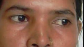
Peripheral vestibular. This common form of nystagmus results from unilateral damage to the vestibular end organ or extra-axial portion of the vestibular nerve. The prototypical cause is acute labyrinthitis or vestibular neuronitis. Because the lesion is strictly unilateral, there will be asymmetric stimulation of the vestibulo-ocular reflex, creating a slow conjugate ocular drift toward the side of the lesion. That drift is met by contraversive quick phases that constitute a jerk nystagmus directed away from the side of the lesion. The input from all three semicircular canals is typically impeded, so the nystagmus will have a horizontal-rotary trajectory. Unlike nystagmus generated by central nervous system lesions, it is “unidirectional.” That is, it always beats away from the side of the lesion regardless of the position of the eyes. Nystagmus amplitude will be highest in far eccentric gaze away from the side of the lesion, intermediate in primary position, and least in eccentric gaze toward the side of the lesion. The nystagmus trajectory will remain in the horizontal plane even in upgaze and downgaze (“uniplanar”), another feature that distinguishes it from the nystagmus of brainstem or cerebellar origin.
Another important feature that distinguishes peripheral from central nystagmus is that visual fixation suppresses it. Nystagmus not apparent under normal viewing conditions can often be drawn out by blurring vision with high-plus (Frenzel) lenses or by performing direct ophthalmoscopy on one eye while shielding the other eye.
Unlike acute vestibular inflammation, the cupulolithiasis that causes benign paroxysmal positional vertigo usually affects only the posterior semicircular canal, creating an upbeat rotary nystagmus toward the dependent ear. Less common BPPV variants may produce horizontal or downbeat nystagmus (20). Head-hanging maneuvers (Dix-Hallpike, Nilen-Barany) are usually necessary to draw out this nystagmus. The nystagmus appears after a latency of several seconds, speeds up, and dissipates within less than 60 seconds. In the less common horizontal canal BPPV, horizontal nystagmus beats towards the ground (“geotropic”) or away from the ground (“apogeotropic”).
Canalith repositioning maneuvers, often very effective in reversing the clinical manifestations of BPPV, are performed based on the putatively involved semicircular canal. For example, the Epley maneuver is used to treat posterior canal BPPV and can be initiated directly from a positive Dix-Hallpike maneuver. The head is rotated 90 degrees away from the affected ear and held in that position for 30 seconds. The head is then rotated another 90 degrees in the same direction and held for 30 seconds before the patient is returned to the sitting position. Canalith repositioning maneuvers sometimes need to be repeated (12).
In acute peripheral vestibular lesions, the head impulse test will be positive, nystagmus will be unidirectional and remain horizontal-rotary in upgaze and downgaze, and there will be no skew deviation (Head Impulse test + nystagmus + test of skew = HINTS) (17). However, the results of the HINTS evaluation are not entirely reliable, so they should be supported by other pertinent distinguishing features, including tinnitus, hearing loss, falling to one side (peripheral lesions), and ataxia (central lesions) (18; 24).
External compression of one side of the pons by a cerebellopontine angle tumor can produce a curious hybrid of gaze-evoked and vestibular nystagmus known as “Bruns nystagmus.” Horizontal gaze toward the side of the lesion elicits a high-amplitude, low frequency, gaze-evoked horizontal jerk nystagmus attributed to impairment of central horizontal gaze-holding centers. Horizontal gaze away from the side of the lesion elicits a small-amplitude, high-frequency, horizontal rotary-jerk nystagmus attributed to unbalanced vestibular tone owing to damage to the eighth nerve.
Prognosis and complications depend on the form and etiology of the nystagmus.
Case 1. A 40-year-old man presented to the office complaining of vertigo. He said that the spinning sensation began suddenly when he turned his head. He also experienced nausea and felt unsteady when he walked. His wife said that he walked as though he were drunk. On examination, the patient’s visual function was normal, including visual acuity, color vision, visual fields, and funduscopic examination. On ocular motility testing, the patient had right-beating nystagmus, which increased in amplitude on looking to the right and reduced in amplitude on looking to the left. The nystagmus became more obvious when one eye was dazzled with bright light while the other eye was intermittently covered. This is an example of an acute left vestibular neuritis.
Case 2. An 18-year-old man presented with complaints of frequent falls over the past 6 months. His brother reported that “he sounds like he has marbles in his mouth.” When asked, the patient reported that his vision was blurry. He had downbeat nystagmus in primary position that increased in down-and-lateral gaze. An MRI of the brain revealed a Chiari malformation with 16 mm downward herniation of the cerebellar tonsils. He underwent an occipital decompression with improvement in his speech and ataxia. There was slight improvement in the nystagmus.
Case 3. A 25-year-old woman presented with headache, diplopia, and difficulty walking of 4 months duration. On examination, in addition to right fifth, sixth, seventh, and eighth cranial nerve palsies, she had marked gait ataxia. The left plantar reflex was extensor. Examination of eye movements revealed a low frequency, coarse right-beating nystagmus on looking to the right and a high frequency, small amplitude, left-beating nystagmus on looking to the left. MRI of the brain showed a large right cerebello-pontine angle tumor. The combination of these two types of nystagmus is characteristic of Bruns nystagmus.
The principal clinical challenges are to distinguish nystagmus from its imitators, physiologic from pathology nystagmus, pendular from jerk nystagmus, and early childhood-onset from later-onset nystagmus.
Careful observation of the characteristics of the nystagmus, the age of the patient, and accompanying non-ocular features form the basis of diagnosis.
The imitators of nystagmus are saccadic intrusions (square wave jerks, macrosaccadic oscillations, ocular flutter, opsoclonus, volitional flutter, and saccades of inattention) as well as other ocular oscillations that do meet the dynamic specifications of nystagmus or saccadic intrusions (convergence retraction, superior oblique myokymia, ocular bobbing, and ping-pong or periodic alternating gaze, myasthenia gravis, and ocular motor palsy) (20).
Saccadic intrusions.
Square wave jerks. This is a common type of saccadic intrusion in which the eyes move arrhythmically and conjugately off to one side of fixation, pause (“intersaccadic interval”), and then return to fixation. If the intrusions are very frequent, the patient may report impaired vision because of interruptions in fixation. Small amplitude (less than 2 degrees) square wave jerks are considered physiologic; larger amplitude square wave jerks are pathologic, occurring especially in progressive supranuclear palsy, recessive ataxias, and after severe closed head injury.
Macrosaccadic oscillations. These oscillations consist of arrhythmic large-amplitude fast phases with an intersaccadic interval. They are stimulated by shifts of fixation that carry the eyes beyond the viewed target, followed by oscillations about the target with decreasing amplitude until the eyes become motionless as they fixate. Macrosaccadic oscillations probably represent a form of ocular dysmetria akin to ataxia, a manifestation of disease of the cerebellar pathways.
Ocular flutter and opsoclonus. These oscillations consist of bursts of rapid saccades of relatively low amplitude and without an intersaccadic interval. If they are confined to the horizontal plane, they are called “ocular flutter”; if they also occur in the vertical or torsional plane, they are called “opsoclonus.” Opsoclonus is probably an extreme form of ocular flutter. As opsoclonus begins to resolve, it often turns into ocular flutter. Common accompaniments include myoclonus, ataxia, and a confusional and severe anxiety state. Disinhibition of brainstem ocular motor pulse generators is believed to be at fault.
There are four causes: (1) post-viral and post-vaccinial encephalitis; (2) paraneoplastic encephalitis; (3) toxic-metabolic syndromes; and (4) idiopathic cause. In children, the common paraneoplastic cause is metastatic neuroblastoma; in adults, it is small-cell lung, breast, or ovarian cancer. Toxins include chlordecone, strychnine, toluene, and organophosphates; metabolic conditions include hyperosmolar coma and pregnancy.
There is a form of ocular flutter that can be brought on volitionally, usually by attempted convergence. The oscillations are of very high frequency and short duration, interrupted by periocular discomfort. They may also be accompanied by blepharospasm and intermittent convergence spasm.
Saccades of inattention. Patients who are inattentive because of distractability, impersistence, or intense concentration on a thought may display wide-amplitude arrhythmic saccades that carry the eyes conjugately off target with long pauses.
Other imitators of nystagmus.
Convergence retraction. On attempted upgaze, all extraocular muscles co-contract, retracting the eye into the orbit. Because the medial rectus muscles are the most powerful extraocular muscles, the eyes also converge. An optokinetic stimulus moved in the downward direction is useful in drawing out this aberrant ocular movement.
Because these convergence retraction movements are not initiated by a slow conjugate ocular drift, they do not represent a true nystagmus but, rather, a form of clonus. They are always accompanied by impaired upgaze, and sometimes lid retraction (“Collier’s sign”), pupillary light-near dissociation, skew deviation, fourth nerve palsy, and comitant esotropia or exotropia.
The primary dysfunction is in the posterior commissure. Lesions in the caudal thalamus, pretectum, dorsal midbrain, or pineal gland are often responsible, but obstructive hydrocephalus from aqueductal stenosis or ventriculoperitoneal shunt failure is frequently implicated (41). Commonly called the “dorsal midbrain syndrome,” this entity is also known as the pretectal syndrome, Parinaud syndrome, and Koeber-Salus-Elschnig syndrome.
Superior oblique myokymia. This condition consists of arrhythmic monocular intorsional movements, usually evoked by downward gaze (14). The patient will report very annoying intermittent diplopia and a “twitching” sensation in the affected eye. The ocular movement is often too subtle to be visualized without magnification, but it can be observed with the slit lamp biomicroscope.
Although the cluster-like episodes often resolve spontaneously, they often recur. The mechanism is believed to be irritability of the extra-axial portion of the fourth nerve. If the myokymia is an isolated abnormality, brain MRI is not indicated. Treatment with topical beta-blockers or systemic anticonvulsants may be effective.
Ocular bobbing and its variants. This eye movement abnormality consists of arrhythmic fast conjugate downward movements followed by a slow return to the primary gaze position. There is an inverse form called “ocular dipping,” in which the eyes move down slowly and come up rapidly. There is also a variant called “reverse bobbing” in which the eyes move up rapidly beyond primary gaze position and come down slowly.
Another variant is called “reverse ocular dipping” in which the eyes move up slowly and come down rapidly. Ocular bobbing is said to indicate severe pontine dysfunction but also toxic metabolism disorders. The other variants have less localizing value, being associated often with diffuse ischemic, metabolic, or toxic brain damage (20). The mechanism may be a release of vertical gaze pathways.
Ping-pong gaze and periodic alternating gaze deviation. In these conditions, the eyes move horizontally from side to side, alternating direction rapidly (ping-pong gaze) or slowly (periodic alternating gaze). Underlying causes are midbrain ischemia, bilateral cerebral ischemia, and hepatic encephalopathy. Although these eye movements are often mistakenly considered to be manifestations of seizures, the electroencephalogram typically shows diffuse slowing, and the patient is in a vegetative state (20). The mechanism may be a release of horizontal ocular motor pathways.
Myasthenia gravis. Myasthenia gravis may exhibit ocular oscillations of two types: (1) brief jerk nystagmus in the extremes of gaze (“gaze-evoked”), most common in the abducting eye and increasing in amplitude with sustained eccentric gaze; it mimics the abducting nystagmus of internuclear ophthalmoplegia; (2) brief high-frequency oscillations in primary position after an attempted saccade (“quiver movements”); they do not occur in any other condition (20).
Ocular motor palsy. Impaired transmission by the third, fourth, or sixth cranial nerves may lead to incomplete ocular ductions with a few beats of jerk nystagmus in the direction of gaze. Alternatively, the eye contralateral to an ocular motor palsy may overshoot in eccentric gaze, just as it does in internuclear ophthalmoplegia.
|
• Step one: is this nystagmus or an imitator? | |
|
• Step two: if nystagmus, is it physiologic or pathologic? | |
|
• Step three: if pathologic, is it pendular or jerk? | |
|
• Step four: is onset in early childhood or later in life? |
If the nystagmus is pendular and is of early childhood onset, choose between monocular, circular, or seesaw nystagmus of diencephalic disease, and spasmus nutans. Brain MRI is indicated.
If the nystagmus is pendular and has its onset later in life, choose between the circular nystagmus of multiple sclerosis or oculopalatal tremor and the convergent nystagmus of oculomasticatory myorhythmia, which is pathognomonic of Whipple disease. Brain MRI is indicated with the understanding that in oculopalatal tremor, the only new abnormality will be in the inferior olive.
If the nystagmus is jerk and has its onset in early childhood, the likely choice is congenital nystagmus. That diagnosis compels the exclusion of an underlying sensory ophthalmic disorder. Brain MRI is not indicated unless there are atypical features, including periodic alternating nystagmus, which has an acquired variant.
If the nystagmus is jerk and has its onset later in life, the challenge is to distinguish central from peripheral (vestibular) causes. Among central causes, the most common is the horizontal gaze-evoked (“side beat”) nystagmus of brainstem or cerebellar origin. Other possibilities are downbeat nystagmus, upbeat nystagmus, pure torsional nystagmus, and periodic alternating nystagmus, all of which localize to the brainstem or cerebellum. Peripheral vestibular nystagmus should be distinguishable by the fact that it is unidirectional and uniplanar and is often accompanied by vertigo, past-pointing, falling to the side of the lesion, and sometimes by auditory features, such as tinnitus and hearing loss. Nystagmus fits into the “HINTS” compendium, which includes the head impulse test and skew deviation.
The treatments directed at reducing nystagmus amplitude, and thereby improving visual acuity or reducing oscillopsia, have included medications, canalith repositioning, optical devices, botulinum toxin injections, and eye muscle surgery. With some exceptions, most of these interventions have been marginally effective, often exacerbating the nystagmus and accompanying neurologic manifestations.
Medications. The potassium channel blocker 4-aminopyridine (dalfampridine 10 mg, twice daily) has effectively reduced downbeat nystagmus amplitude but has caused insomnia, dizziness, headache, paresthesias, and has induced seizures in patients with epilepsy (34; 20). In a double-masked study, neither clonazepam nor baclofen were effective. Anticholinergic agents, such as scopolamine, are also unhelpful and likely to exacerbate accompanying neurologic manifestations. Memantine and dalfampridine may be marginally effective in upbeat nystagmus.
Periodic alternating nystagmus may be responsive to baclofen or memantine (16). The acquired pendular nystagmus of multiple sclerosis and oculopalatal tremor may be responsive to gabapentin (04; 03) or memantine (33). Clonazepam may be effective in seesaw nystagmus and memantine in hemi-seesaw nystagmus (21; 34; 22).
The nystagmus of acute peripheral vestibular dysfunction typically resolves within days so that no treatment for nystagmus is needed.
Canalith repositioning maneuvers. These are often highly effective for the treatment of benign paroxysmal positional vertigo (12).
Optical devices. Patients with congenital nystagmus whose nystagmus amplitude is reduced by convergence may gain slight improvement in visual acuity from spectacle prisms that elicit convergence even as the patient fixates distant targets. High-plus spectacles combined with high-minus contact lenses, which are aimed at stabilizing images on the retina, have been only slightly helpful in reducing oscillopsia and visual acuity in patients with acquired pendular nystagmus (32).
Botulinum toxin injection. Injection of botulinum toxin into the extraocular muscles or retrobulbar space has not been effective because it cripples eye movements and causes diplopia and ptosis (19).
Extraocular muscle surgery. Extraocular muscle surgery has been effective in improving visual acuity in patients with congenital nystagmus who have a face turn and improved visual acuity when their eyes are placed within the null zone (30). The artificial divergence procedure of Cuppers has been helpful in some patients whose congenital nystagmus lessens with convergence.
All contributors' financial relationships have been reviewed and mitigated to ensure that this and every other article is free from commercial bias.

Jonathan D Trobe MD
Dr. Trobe of the University of Michigan has no relevant financial relationships to disclose.
See ProfileNearly 3,000 illustrations, including video clips of neurologic disorders.
Every article is reviewed by our esteemed Editorial Board for accuracy and currency.
Full spectrum of neurology in 1,200 comprehensive articles.
Listen to MedLink on the go with Audio versions of each article.
MedLink, LLC
3525 Del Mar Heights Rd, Ste 304
San Diego, CA 92130-2122
Toll Free (U.S. + Canada): 800-452-2400
US Number: +1-619-640-4660
Support: service@medlink.com
Editor: editor@medlink.com
ISSN: 2831-9125
Neurobehavioral & Cognitive Disorders
Jun. 17, 2026
Neuro-Oncology
May. 27, 2026
Neuro-Oncology
May. 27, 2026
Neuropharmacology & Neurotherapeutics
May. 14, 2026
General Child Neurology
May. 12, 2026
Neuro-Oncology
Apr. 30, 2026
Neuropharmacology & Neurotherapeutics
Apr. 23, 2026
Neuropharmacology & Neurotherapeutics
Apr. 20, 2026