General Neurology
Principles of rehabilitation of patients with neurologic disorders
Oct. 07, 2023
MedLink®, LLC
3525 Del Mar Heights Rd, Ste 304
San Diego, CA 92130-2122
Toll Free (U.S. + Canada): 800-452-2400
US Number: +1-619-640-4660
Support: service@medlink.com
Editor: editor@medlink.com
ISSN: 2831-9125
Toll Free (U.S. + Canada): 800-452-2400
US Number: +1-619-640-4660
Support: service@medlink.com
Editor: editor@medlink.com
ISSN: 2831-9125
Nearly 3,000 illustrations, including video clips of neurologic disorders.
Every article is reviewed by our esteemed Editorial Board for accuracy and currency.
Full spectrum of neurology in 1,200 comprehensive articles.
Listen to MedLink on the go with Audio versions of each article.
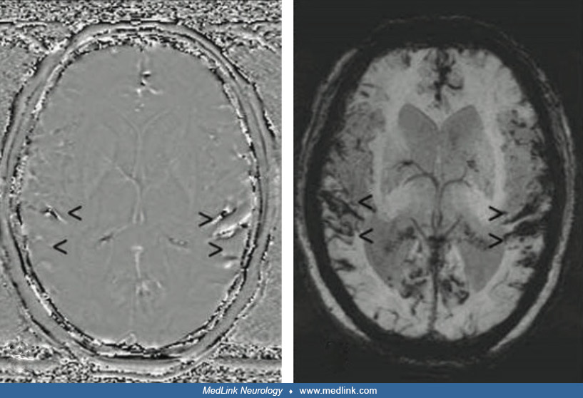
Typical MRI findings in superficial intraventricular surface siderosis. T2-weighted gradient echo (GRE) image shows hemosiderin deposits along the frontal horns (black arrow). (Source: Harizi E, Shemsi K, Ahmetgjekaj I, et al. Superficial intraventricular surface siderosis brain. Radiol Case Rep 2022;17[11]:4152-5. Creative Commons Attribution 4.0 International [CC BY 4.0] license, creativecommons.org/licenses/by/4.0.)