Stroke & Vascular Disorders
Primary prevention of stroke
Dec. 28, 2023
MedLink®, LLC
3525 Del Mar Heights Rd, Ste 304
San Diego, CA 92130-2122
Toll Free (U.S. + Canada): 800-452-2400
US Number: +1-619-640-4660
Support: service@medlink.com
Editor: editor@medlink.com
ISSN: 2831-9125
Toll Free (U.S. + Canada): 800-452-2400
US Number: +1-619-640-4660
Support: service@medlink.com
Editor: editor@medlink.com
ISSN: 2831-9125
Nearly 3,000 illustrations, including video clips of neurologic disorders.
Every article is reviewed by our esteemed Editorial Board for accuracy and currency.
Full spectrum of neurology in 1,200 comprehensive articles.
Listen to MedLink on the go with Audio versions of each article.
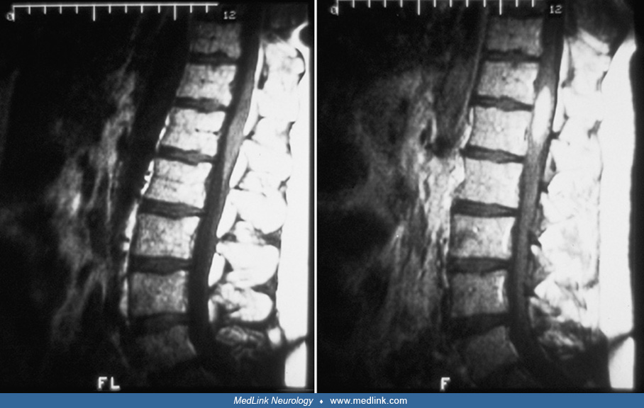
(Left) Unenhanced, sagittal, T1-weighted MRI at the thoracolumbar junction. A metastatic tumor within the conus medullaris is isointense to cord. Above and below the tumor, a thin, tapered, hypointense area represents a syringomyelic cavity or necrotic spinal cord tissue. (Center) Unenhanced, sagittal, T2-weighted MRI. The tumor remains nearly isointense to cord. The surrounding necrotic tissue or syrinx becomes hyperintense. (Right) After the intravenous administration of contrast, a sagittal, T1-weighted MRI shows homogeneous enhancement of the tumor but not the syrinx or necrotic tissue above and below it. (Courtesy of Dr. Judith E Simon.)