Neuropharmacology & Neurotherapeutics
Clobazam
Jan. 15, 2026
MedLink, LLC
3525 Del Mar Heights Rd, Ste 304
San Diego, CA 92130-2122
Toll Free (U.S. + Canada): 800-452-2400
US Number: +1-619-640-4660
Support: service@medlink.com
Editor: editor@medlink.com
ISSN: 2831-9125
Toll Free (U.S. + Canada): 800-452-2400
US Number: +1-619-640-4660
Support: service@medlink.com
Editor: editor@medlink.com
ISSN: 2831-9125
Nearly 3,000 illustrations, including video clips of neurologic disorders.
Every article is reviewed by our esteemed Editorial Board for accuracy and currency.
Full spectrum of neurology in 1,200 comprehensive articles.
Listen to MedLink on the go with Audio versions of each article.
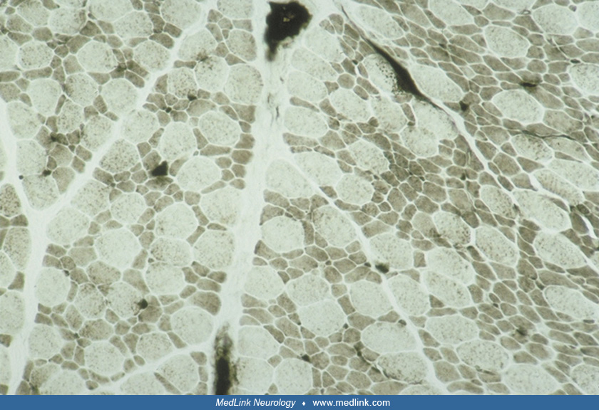
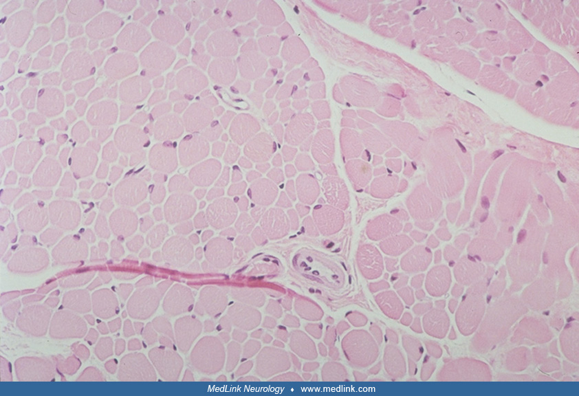
This muscle biopsy is from a 6-month-old boy with congenital muscle fiber-type disproportion. Formalin-fixed, paraffin-embedded, transverse section stained with hematoxylin-eosin shows normal fascicular architecture of the muscle, but 2 populations of fibers by size, hypoplastic and normal to hypertrophic fibers, neither of which exhibit central nuclei or other cytoarchitectural alterations; there is absence of myofiber necrosis, regeneration, or inflammation. Magnification x 100. (Contributed by Dr. Harvey Sarnat.)