Stroke & Vascular Disorders
Carotid-cavernous fistulas
Feb. 03, 2026
MedLink, LLC
3525 Del Mar Heights Rd, Ste 304
San Diego, CA 92130-2122
Toll Free (U.S. + Canada): 800-452-2400
US Number: +1-619-640-4660
Support: service@medlink.com
Editor: editor@medlink.com
ISSN: 2831-9125
Toll Free (U.S. + Canada): 800-452-2400
US Number: +1-619-640-4660
Support: service@medlink.com
Editor: editor@medlink.com
ISSN: 2831-9125
Nearly 3,000 illustrations, including video clips of neurologic disorders.
Every article is reviewed by our esteemed Editorial Board for accuracy and currency.
Full spectrum of neurology in 1,200 comprehensive articles.
Listen to MedLink on the go with Audio versions of each article.
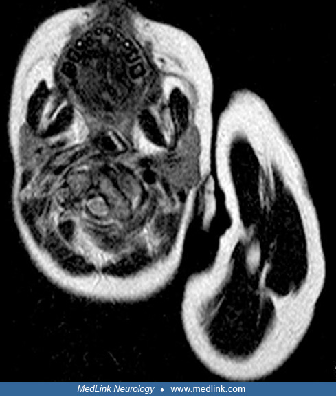
Axial CT through the ring of C1 and the body of C2 showing fixed rotatory dislocation due to bilateral facet subluxation. The right C1 facet has subluxed anteriorly with respect to the right C1 facet. The left C1 facet has subluxed posteriorly with respect to the left C1 facet. The patient’s head would be turned to the left and tilted to the right. Note that the spinal canal is diminished in diameter. The ring of C1 has moved forward and rotated to the left. The dens of C2 appears close to the right C1 lateral mass. Note that the spinal canal remains capacious, without imminent spinal cord compression. “Od” refers to the odontoid process. “VA” is adjacent to the left foramen transversarium of the ring of C1, through which the vertebral artery passes. (Contributed by Dr. David Choi.)