Neurobehavioral & Cognitive Disorders
Mental status examination
Jun. 17, 2026
MedLink, LLC
3525 Del Mar Heights Rd, Ste 304
San Diego, CA 92130-2122
Toll Free (U.S. + Canada): 800-452-2400
US Number: +1-619-640-4660
Support: service@medlink.com
Editor: editor@medlink.com
ISSN: 2831-9125
Toll Free (U.S. + Canada): 800-452-2400
US Number: +1-619-640-4660
Support: service@medlink.com
Editor: editor@medlink.com
ISSN: 2831-9125
Worddefinition
At vero eos et accusamus et iusto odio dignissimos ducimus qui blanditiis praesentium voluptatum deleniti atque corrupti quos dolores et quas.
Electrical injury can affect any organ. Neurologic complications are well-recognized and cause a distinct increase in morbidity. The broad range of neurologic complications of electrical injuries are described, including peripheral neuropathy, central nervous system disorders, and seizures. Clinical features that are useful in evaluating patients after electrical injury are identified. Neurologic sequelae of electrical injuries may present years after electrical injury. Neuropsychological disorders may present as posttraumatic stress disorder. The management of late neurologic sequelae of electrical injury is described.
Note that a separate MedLink article addresses Lightning injuries: neurologic complications.
• Neurologic sequelae from electrical trauma can be immediate (transient or permanent), delayed and progressive, or linked to a secondary injury caused by electrical injury. | |
• There is an increase in the incidence of neuropsychiatric sequelae of electrical injury, such as posttraumatic stress disorder, the second most common type of sequelae after those related to burns. | |
• Neurologic sequelae include loss of consciousness, headache, memory disturbance, seizures, movement disorders, speech impediment and mutism, vertigo, cranial nerve dysfunction, meningitis, autonomic nervous system complications, and peripheral neuropathies. | |
• The diagnostic approach to the neurologic consequences of an electrical injury patient is like the neurologic evaluation of a multi-trauma patient. | |
• Indications for hospital admission include the following: exposure to an electrical source of more than 200 V and 200 mA; cardiac abnormalities; loss of consciousness; fall from a height; thermal injury to greater than 15% of the body surface area or burns to the hands, feet, face, or groin; respiratory distress; spine fractures; serum electrolyte derangements; and compartment syndromes. |
The scientific study of electricity developed in the 17th and, particularly, 18th centuries, as did the study of electrical effects on human and animal bodies.
A Leyden jar (or Leiden jar, from archaic Dutch, or sometimes Kleistian jar) is an electrical device that stores a high-voltage electric charge between electrical conductors on the inside and outside of a glass jar--the original form of the capacitor or condenser. It was invented, apparently independently, by German cleric Ewald Georg von Kleist (c. 1700-1748) in 1745 and by Dutch scientist Pieter van Musschenbroek of Leiden (Leyden), Netherlands c1745-1746 (06) and his student Andreas Cunaeus. Musschenbroek and Cunaeus each received strong shocks from the capacitance discharge when they attempted to "condense" electricity in a glass of water.
Dutch scientist Pieter van Musschenbroek of Leiden (Leyden), Netherlands c1745-1746 (Allerhand 2018) and his student Andreas Cunaeus performing electrical experiments. (Source: Print from a steel engraving by Christiaan Lodewij...
|
I hung a copper wire ... from the end of the iron tube, and ... I took a glass vase from which we are wont to drink beer. This [glass] was filled half with water. Then the wire was hung in the middle of the water. When the [glass] globe was rotated [to generate a static charge], such heavy sparks were drawn from the wire to the finger that the pain was unbearable, and even the vase itself got cracks. The water remained electric for a long time and gave repeated explosions to the finger ... (from Musschenbroek 1745, translated by Present in 2022, page 118) (64). |
Ultimately, before other means were developed to provide electricity, banks of Leyden jars were employed to store large-voltage electric charge for such things as wireless communications (ie, the telegraph).
Spark-gap radio transmitter for a long-distance wireless telegraphy radio station built by Gesellshaft fur Drahtlose Telegraphie (Telefunken) at Nauen, Germany in 1907. Designed by German radio pioneer Georg von Arco (1869-1940...
American statesman and scientist Benjamin Franklin (1706-1790) was the first to point out the relation of lightning to electricity in 1752. Franklin later invented the lightning rod and, in an infamous episode, attempted to use electricity to electrocute a turkey.
Detail of the engraving of Benjamin Franklin by James McArdell after a painting by Benjamin Wilson. The image has been rotated to allow easy readability. (Courtesy of the U.S. Library of Congress, Washington, D.C. Public domain...
Italian physician, anatomist, and physiologist Luigi Galvani (1737-1798) established the existence of what he called “animal electricity” (animalis electricitas) (26; 59; 60; 61; 13; 44).
After more than a decade of research, Galvani described his results in De Viribus Electricitatis in Motu Musculari Commentarius (Commentary on the Effects of Electricity on Muscular Motion) (29). This was reprinted at Modena, 1792, with a note and dissertation by Galvani's nephew, Giovanni Aldini (1762-1834), and with new plates (30).
Galvani's experiments with static electricity transmitted to the spinal cords of decapitated frogs. Some of the frog legs are open to the air, and others are sealed in jars but still connected to wires passing through a cork at...
Although various individuals had previously suggested that electricity played a role in nervous action, Galvani’s experimental results garnered international attention. Although some derided Galvani’s notions, many considered to have proven both the existence of animal electricity and the operation of this electricity through nerves to produce muscular contractions.
Italian physicist Alessandro Volta (1745-1827) was among the first scientists to repeat Galvani’s experiments.
Initially, Volta enthusiastically supported Galvani's work, but he soon began to doubt that the observed muscle contractions were due to intrinsic electricity in animals. Volta suggested instead that the muscle contractions were due to an extrinsic form of electricity that was simply put in motion through the conducting metals or created by dissimilar metals.
From 1792 to 1797, German polymath Alexander von Humboldt (1769-1859) replicated many of the experiments of Galvani and Volta.
Humboldt concluded that although the interaction of dissimilar metals could be an effective stimulus for muscle contraction (as both Galvani and Volta agreed), Galvani was nevertheless correct in believing that this was not necessary for muscles to contract and, further, that the preponderance of evidence supported the existence of an intrinsic form of “animal electricity.” However, Humboldt was not convinced that Galvani’s “animal electricity” was equivalent with other forms of electricity.
By 1800, Volta had constructed electrical batteries consisting of two different metals in an electrolytic salt solution. In addition, Volta discovered that a much greater electrical effect could be produced by a series of alternating copper and zinc disks placed in a line, with each copper-zinc pair separated from the next pair by a cloth or cardboard disk soaked in brine. This device, which Volta called a “pile” and is consequently now referred to as a “voltaic pile,” was the first practical battery.
Throughout the scientific controversy, Galvani was unwilling to engage directly in arguments against the challenges of Volta and others. Consequently, Galvani's nephew Giovanni Aldini became the main defender and proponent of Galvani’s concept of animal electricity (55; 44; 09).
Aldini traveled across Europe, publicly electrifying human and animal bodies in theatrical performances. In 1802, Aldini went to London and elicited repeated, spasmodic movements of facial muscles on human heads, arms, and legs as well as the heads and trunks of oxen, cows, horses, sheep, and dogs. Aldini's most famous performance was for a large medical and general audience at the Royal College of Surgeons in London in 1803 on a hanged man named George Forster, who had been convicted of murdering his wife and child by drowning them in the Paddington Canal. Aldini took a pair of conducting rods linked to a powerful voltaic pile and touched the rods to various parts of the corpse. When the rods were applied to Forster’s mouth and ear, “the jaw began to quiver, the adjoining muscles were horribly contorted, and the left eye actually opened” (04). When one rod was moved to touch the rectum, the whole body convulsed, and the movements were “so much increased as almost to give an appearance of re-animation” (04).
Leyden jars and other apparatus used by Aldini in experiments, which were intended to support his uncle, Galvani, against the attacks of Volta. (Source: Plate 4. Aldini J. An account of the late improvements in galvanism, with ...
Aldini's Essai théorique et expérimental sur le galvanisme: avec une série d'expériences faites en présence des commissaires de l'Institut National de France, et en divers amphithéâtres anatomiques de Londres ... [Theoretical and Experimental Essay on Galvanism: With a Series of Experiments Made in the Presence of the Commissioners of the National Institute of France, and in Various Anatomical Amphitheaters in London] contained a full account of his various “experiments” as well as numerous engravings (05). A decade later, Aldini's spectacles served as an inspiration for Mary Shelley's novel Frankenstein; or, The Modern Prometheus (1818) and for various political cartoons.
Electric motors were invented in the early part of the 19th century (1820s-1830s), and with the development of power stations and electrical lightning, electricity entered the industry and daily life of people. In 1882, Thomas Edison (1847–1931) formed the Edison Electric Illuminating Company of New York, bringing electric light to parts of Manhattan. Nevertheless, most Americans still lit their homes with gas lights and candles for another 50 years.
One bizarre electrocution event of an elephant in 1903 has erroneously been attributed to an anti-alternating current demonstration organized by Thomas A Edison (who favored direct current) during the so-called "war of the currents." Topsy (c 1875 – January 4, 1903) was a female Asian elephant who was electrocuted at Coney Island, New York, in January 1903. During her 25 years at the Forepaugh Circus, Topsy gained a reputation as a "bad" elephant and, after killing a spectator in 1902, was sold to Coney Island's Sea Lion Park. During the construction of the park that took its place, Luna Park, Topsy was used in publicity stunts. Luna Park's publicity-hungry owners, Frederic Thompson and Elmer "Skip" Dundy planned to advertise the opening of their new park by euthanizing Topsy in a public hanging and charging admission to see the spectacle, but this was prevented by the American Society for the Prevention of Cruelty to Animals. The event was instead limited to invited guests and press only. On January 4, 1903, in front of a small crowd of invited reporters and guests, Topsy was fed carrots laced with 460 grams of potassium cyanide, electrocuted and strangled, the electrocution being the final cause of death. Among the invited press that day was a crew from Edison Studios who filmed the event. Their film of the electrocution was released to be viewed in coin-operated kinetoscopes under the title "Electrocuting an Elephant." Although Edison Studios filmed the event, Edison himself was never at Luna Park, and the electrocution of Topsy took place 10 years after the war of currents.
Serbian-American inventor and electrical engineer Nikola Tesla (1856–1943) is known for his contributions to the design of the modern alternating current (AC) electricity supply system.
A drawing of Serbian-American electrical engineer Nikola Tesla illuminated by electrical discharges created by touching the high voltage terminal of his Tesla coil oscillator, from an illustration accompanying an 1894 interview...
Tesla emigrated to the United States in 1884 and began working almost immediately at the overcrowded Edison Machine Works on Manhattan's Lower East Side. Tesla quit after 6 months. In late 1886, after several other failed endeavors, the Tesla Electric Company was formed in April 1887 with the assistance of venture capitalists. In 1887, Tesla developed an induction motor that ran on alternating current (AC), a power system format that was rapidly expanding in Europe and the United States because of its advantages in long-distance, high-voltage transmission. The motor used polyphase current, which generated a rotating magnetic field to turn the motor (a principle that Tesla claimed to have conceived in 1882). This innovative electric motor, patented in May 1888, was a simple self-starting design that did not need a commutator, thus, avoiding sparking and the high maintenance of constantly servicing and replacing mechanical brushes.
Engineers working for the Westinghouse Electric & Manufacturing Company reported to George Westinghouse (1846-1914) that Tesla had a viable AC motor and related power system—something Westinghouse needed for the alternating current system he was already marketing.
In July 1888, a licensing deal was negotiated with George Westinghouse for Tesla's polyphase induction motor and transformer designs. Westinghouse also hired Tesla for to be a consultant at the Westinghouse Electric & Manufacturing Company's Pittsburgh labs 1 year.
It was not until 1925 that half of all homes in the United States had electric power. Most of the earliest knowledge of electrical injury in this period was based on anecdotal reports.
Scientific characterization of human response to electricity did not progress significantly until the last half of the 20th century and was first thoroughly documented in a special report (21). Injuries from exposure to electricity may be superficial, resulting in skin burns if no electrical current travels through the body. Nevertheless, a patient who presents initially with electrical burns and no neurologic signs may develop late neurologic sequelae. In true electrical injuries, the victim becomes part of the electrical circuit with an entrance and exit site for the current. This can involve damage to various organs, including the nervous system. Secondary injuries may be due to falls caused by contact with electrical energy.
Most electrical injuries occur in workers dealing with electric machinery or power lines. The incidence of electrical injuries has decreased with the regulation of occupational electrical safety standards. Electrical injuries can occur at home due to mishaps in handling electric appliances.
Fatal electrical shock is referred to as electrocution, although the term "electrocution" is often used erroneously to refer to nonfatal electrical injuries. The earliest electrocution occurred in a theater in 1879, whereas intentional electrical fatality as a form of capital punishment dates to the first electric chair execution in 1890 (83).
According to the American National Standards Institute criteria (ANSI C84.1-1989), system voltages are categorized by “voltage classes.” Voltages 600 V and below are referred to as “low voltage." Such voltages can still produce serious neurologic injuries or even death, but they are officially classed as “low voltage."
|
• The medical manifestations of electrical injuries may evolve over minutes to hours and days. | |
|
• Delayed sequelae may occur years following an electrical exposure. |
Electrical injuries are associated with multiple sequelae. Immediate complications include cardiac and renal injuries, as well as deep burns (40). Neurologic complications, although relatively uncommon, can occur immediately or may be delayed (40).
Categories of neurologic sequelae that may result from electrical injuries include the following:
|
• immediate and transient, in which symptoms occur at the time of the incident and remit within hours to days (eg, loss of consciousness or retrograde amnesia); | |
|
• immediate and prolonged or permanent, in which symptoms occur at the time of the incident and persist for weeks, months, years, or indefinitely (eg, brain hematoma or infarction); | |
|
• delayed and progressive, in which symptoms were not present at the time of the incident but appear at some later time (days, months, or even years), and the severity of the symptoms progresses (eg, movement disorders, demyelinating disorders, and cerebrovascular occlusive disease); and | |
|
• linked or coupled, when electrical injury causes secondary injury (eg, falls and head injury). |
In nonfatal injuries, the clinical course can be variable. Opinions differ about the nature and cause of patient symptoms and the relationship between symptoms and factors like trauma severity, litigation, or premorbid personality. Not all survivors develop physical, cognitive, or emotional difficulties. No consistent relationship has been established between characteristics such as age, injury-related characteristics (eg, voltage, current source, work error), and neuropsychological test performance. Low-voltage electrical injuries usually produce more frequent long-term sequelae than high-voltage injuries.
Neurologic sequelae. Neurologic sequelae of electrical injury are diverse and can include loss of consciousness, confusion or encephalopathy, memory disturbance, seizures, cerebrovascular complications (eg, cerebral hemorrhage, cerebral ischemia, and cerebral venous thrombosis), cerebral edema, hydrocephalus (01), movement disorders (eg, tremors, dystonia, myoclonus, parkinsonism, choreoathetosis, and myokymia), speech impediment or mutism (24; 51), headache, neuro-otological problems (eg, sensorineural hearing loss, tinnitus, dizziness, and vertigo) (53; 85), facial palsy and other cranial nerve dysfunction (02; 65), meningitis, myelopathy, autonomic nervous system complications (eg, reflex sympathetic dystrophy or complex regional pain syndrome), peripheral neuropathies (84), various muscular symptoms (eg, muscle aches, spasm, tetany or cramps, and twitches), and rhabdomyolysis (39).
Some uncommon neurologic electrical injuries include the following:
|
• Rupture of pre-existing vascular malformations (28) | |
|
• Rupture of the carotid artery (79) | |
|
• Development of an intracranial arteriovenous fistula (41) | |
|
• Ischemic stroke in the vertebrobasilar territory as a delayed sequel of electrical injury (37) | |
|
• Reversible bulbar dysfunction with negative MRI following high-voltage occupational electrical injury (57) | |
|
• Right facial palsy and hemotympanum in a 3-year-old boy (65) | |
|
• Flaccid tetraplegia due to very severe generalized sensory-motor polyneuropathy from an intentional high-voltage electrical injury, with subsequent remarkable reinnervation and unmasking of a myelopathy (78) | |
|
• Brachial plexopathy with partial Horner syndrome and phrenic nerve palsy (70) | |
|
• Delayed compressive peripheral neuropathy caused by scarring from cutaneous electrical burns | |
|
• Carpal tunnel syndrome in the absence of severe scarring likely caused by direct nerve injury (84) |
In a study on patients with mild cognitive impairment following electrical injury, cerebral blood volume maps showed hypermetabolism in the cerebello-limbic system, but diffuse tensor imaging did not identify any microstructural changes (56).
Electrical burns in adults are mainly low-voltage burns contracted by manual workers resulting in a flesh burn. In a retrospective study of 82 adult patients with electrical burns admitted to a tertiary burn center, the mean age was 40 years and 93% were men (03). The most common activities causing the injuries were work (39%) and do-it-yourself activities (32%). A low-voltage power source was involved in 78% of cases. The average total body surface area involved was 3%. The most frequently injured body parts were the head, hands, and non-hand areas of the upper extremities. The mean hospital stay was 2 days. Minor injuries were successfully managed nonoperatively. Although rare, the loss of digits, neurologic sequelae, cardiac arrhythmias, and renal failure are serious potential complications.
In another retrospective study of 162 patients with electrical injuries treated at a burn center, 55 suffered low-voltage (< 1000 volts), 55 high-voltage (> 1000 volts), and 52 unknown-voltage injuries (40). Subjects with high-voltage injuries were more likely to be male, experience loss of consciousness, suffer cardiac arrest, and undergo amputation. No significant differences were observed in long-term neurologic deficits. Twenty-seven patients (17%) were found to have neurologic deficits on or after admission; 48% recovered, 33% persisted, 7% died, and 11% did not follow up.
Neuro-urological electrical injuries. Neuro-urological electrical injuries may occur secondary to brain or spinal cord trauma or as independent consequences of electrical shock (86). The main urological functions that can be affected by an electrical injury are lower urinary tract function, erectile function, and renal function:
|
1. Lower urinary tract symptoms following electrical injury should be evaluated clinically, and diagnostic studies (urodynamic and imaging studies) should follow. | ||
|
a. Overactive bladder and detrusor sphincter dyssynergia is the result of the dyssynergy between the detrusor muscle and the urethral sphincter, which can be continuous or intermittent and can be responsible for lower or upper urinary tract infections, upper urinary tract dilatation, and renal failure. Suprapontine cerebral lesions or cervical and thoracic myelopathy secondary to electrical injury can induce this syndrome. | ||
|
b. Underactive bladder appears with urinary retention and voiding dysfunction and can also induce urological complications. Cerebral or spinal lesions between the pontine micturition center in the brainstem and the sacral micturition center in the sacral spinal cord are the usual causes. An underactive bladder is the most common sequel of myelopathy secondary to electrical injury. | ||
|
2. Erectile dysfunction | ||
|
a. Due to high-voltage injuries without recognized neurologic injury (severe penile trauma, penile amputation). | ||
|
b. Due to spinal cord injury. Myelopathy after high-voltage electrical injury or lightning injury can occur as a direct injury at any spinal cord level (73), and it can also occur secondary to indirect spinal cord damage such as spinal fractures due to fall, mainly of the thoracic vertebrae. Renal electrical injury requiring hemofiltration is associated with a high mortality. | ||
Neuropsychological symptoms. These may include memory disturbance, increased emotional sensitivity, insomnia, unusual anxiety, reduced attention span/loss of concentration, personality changes, fear of electricity, and inability to cope. Some neuropsychological symptoms are compatible with posttraumatic stress disorder, which had an estimated prevalence of 7.6% in a retrospective study of 311 cases of occupational electrical injuries (62). Posttraumatic stress disorder is characterized by intrusive thoughts, nightmares and flashbacks of past traumatic events, avoidance of reminders of trauma, hypervigilance, and sleep disturbance, all of which lead to considerable social, occupational, and interpersonal dysfunction. A diagnosis of posttraumatic stress disorder is made for patients older than 6 years of age who meet all the DSM-5 criteria for the syndrome (07).
Nonelectrical effects. With higher power density and smaller, more compact spaces, there is an increased risk of damage from explosive effects with characteristics of closed blast injury.
Blast can result in tissue destruction from shrapnel, confined space air contamination, acoustic or pressure effects, and acceleration-deceleration forces. Traumatic brain injury can occur despite an intact skull. When a patient’s neurologic presentation is consistent with blunt force or blast injury after an electrical event, consideration should be given to the possibility of exposure to an electrical explosion scenario complicating an electrical shock.
Survival from electrical injury can be associated with significant functional impairment. In a landmark retrospective study of employees of a national electrical energy company, the electrical trauma survival experience from 1970 to 1989 was reviewed for a workforce between 100,000 to 120,000 persons (31). Electrical injuries affected 2080 workers. Of these, 515 patients, or 25%, had postinjury complications, including burn-related complications (63%), neuropsychiatric complications (18%), sensory impairment (12%), amputations (5%), orthopedic injury (5%), and cardiovascular impairment (1%).
Sense organ disorders included anosmia; vision-related changes due to conjunctivitis, keratitis, and cataracts; and neuro-otological sequelae with conductive or sensorineural hearing loss, tinnitus, and vertigo. In 59 of the 515 patients, disability was considered serious, with an impairment rating from 31% to 100% (31).
Neurologic complications of electrical injury. Acute CNS complications are well-recognized and cause an increased risk of morbidity, whereas peripheral nervous system complications are less predictable after electrical injuries.
Delayed neurologic sequelae include hydrocephalus, cerebral venous thrombosis, and amyotrophic lateral sclerosis and usually carry a great risk for permanent disability (86). However, a case of acute-onset quadriplegia following a high-voltage electrical injury, with no radiographic abnormalities, regained partial sensorimotor function 2 months after the injury (23).
Cases of delayed onset of visual symptoms have been reported in whom MRI showed bilaterally symmetrical diffusion restriction in the parietal and occipital cortex, and improvement followed intravenous steroids (19; 54).
Osteoporosis may develop as a complication of electrical skin burns and is explained by prolonged sympathetic nervous system dysfunction, which may result in bone metabolism derangement even after the acute phase of electrical burn (67). Other contributing factors include prolonged bed rest and immobilization, as often occurs with severe burns or other injuries.
Measures of physical morbidity, as measured by total body surface burned, total days in intensive care, and hospital length of stay, have been traditionally used as predictors of outcome in high-voltage electrical injuries, but functional outcomes, including neuropsychological sequelae, are similar between disparate levels of electrical injury as judged by voltage (20). Electrical injuries may incur severe morbidity despite relatively small burn size or low voltage (20).
|
• Electrical injuries may occur from accidents during recreational activities or at work, household electric appliances, during medical procedures, and use of weapons such as Taser. | |
|
• Injury involves energy transfer to an exposed individual in some combination of electrical, thermal, radiation, acoustic, and mechanical forms. | |
|
•Late neurologic sequelae are difficult to explain, but several hypotheses have been proposed. |
Etiology. The severity of an electrical injury is determined by many factors, including the type of current, the pathway of the current through the body, the resistance of tissues along this pathway, the voltage and amperage of the current, the duration of contact, and environmental factors, such as weather and humidity (48).
Electrical injuries are categorized into four main types based on how electricity affects the body of an individual, of which only the last is the primary focus of this article (87):
(1) Flash injuries from an arc flash typically cause cutaneous burns, as no electrical current passes through the body.
(2) Flame injuries occur when an arc flash ignites an individual's clothing, leading to thermal burns.
(3) Lightning injuries involve extremely short-duration, high-voltage electrical energy and are characterized by flash-over currents along the skin surface, with variable conduction through the body.
(4) True electrical injuries occur when the individual becomes part of an electrical circuit, usually with identifiable contact points at the entrance and exit sites.
Table 1 highlights nonoccupational causes of electrical injury.
|
Circumstance |
Source |
Examples |
|
Household accident |
Electrical appliances |
Young children inserting metal objects into electrical sockets |
|
Home electric circuits |
Faulty wiring or electrical repairs performed by unqualified people | |
|
Damaged electric cords |
Badly deteriorated electrical cords, plugs, and appliances | |
|
Recreational accident |
Electric devices |
Contact of electrical devices with water |
|
Power lines |
Contact with overhead power lines | |
|
Medical |
Iatrogenic |
Electroconvulsive therapy |
|
Weapon |
Conducted electrical weapon (CEW; eg, stun gun or Taser) |
Use by police to subdue suspected criminals, suspects, and mentally ill subjects who are combative, violent, or suicidal. |
|
Suicide |
Electrical appliance |
Electrical appliance placed in a bathtub |
|
Homicide |
Varies |
Rare but sometimes concealed as apparent suicide (17; 38; 49) |
|
Torture |
Electrodes |
Detainees subjected to torture and extrajudicial execution by State actors and terrorists (63) |
|
Execution |
Electric chair |
Judicial execution* |
|
| ||
Conducted electrical weapons (CEWs). Stun guns and Tasers have been developed for use by law enforcement and security personnel to provide less lethal alternatives to conventional weapons. These devices deliver bursts of high-voltage, low-amperage direct current, either through a handheld device (stun gun) or through a hook-and-wire system fired using compressed gas (Taser). CEWs are widely available and have been used for criminal purposes and torture. The authors of two systematic reviews concluded that prolonged observation and diagnostic testing are not necessary in patients who are otherwise asymptomatic and alert following such an exposure (58; 80). However, in rare instances, various adverse outcomes related to the use of electrical weapons have been described. Examples include cardiac arrhythmias resulting from fatal, blunt, or penetrating injuries, and neurologic sequelae such as seizures, altered mental status, and ischemic stroke (11). With 16 case reports in the literature, the use of Taser presents a small but real risk of death from fatal traumatic brain injury due to a fall, and old age represents an independent risk factor for such fatalities (42).
Electroconvulsive therapy (ECT). ECT is still used for the treatment of some psychiatric disorders and typically causes some amnesia (18) or headaches (68) and rarely may produce other neurologic disturbances. Usually, the peri-event amnesia and neurologic complications of ECT are transient, but this is not universally true. Headaches are usually mild and short-lasting, but recurrent common migraine has been reported (68).
As argued by proponents of ectroconvulsive therapy (25):
|
• Prospective neuroimaging studies have shown no evidence of ECT-induced structural changes. | ||
|
• Early human autopsy cases with cerebrovascular lesions after ECT are now discounted as purportedly due to agonal changes or undiagnosed preexisting disease. | ||
|
• In animal studies that used a stimulus intensity and frequency comparable to human ECT, no neuronal loss was seen when appropriate controls and blind ratings were employed. | ||
|
• Neurologic and psychiatric complications have been reported in the modern era following ECT; some recent cases and studies are summarized below: | ||
|
- A 46-year-old depressed woman treated with four bilateral, bitemporal ECT procedures developed delusions of reference, persecution, agitation, oneiric delusions, and olfactory hallucinations after the fourth ECT session. These persisted for 14 days but resolved after treatment with olanzapine (08). | ||
|
- In a retrospective study of 295 patients undergoing ECT, severe postictal confusion occurred in 25%, and no clear risk factors emerged from a multivariate analysis (71). | ||
|
- A systematic review identified 39 cases with modified ECT-related tardive seizures (81). Almost all had at least one identified risk factor for seizures, though none were ultimately diagnosed with epilepsy even if 20% continued on antiepileptic medications. | ||
|
- Five patients developed temporal epileptiform abnormalities on EEG after receiving maintenance ECT for at least 8 months, despite no history of epilepsy and no abnormality on neuroimaging (16). EEGs normalized in these cases after cessation of ECT, and there were no further clinical seizures. | ||
|
- An 18-year-old woman with paranoid schizophrenia experienced cortical blindness immediately after her first bifrontotemporal ECT treatment but spontaneously recovered after 6 hours (76). | ||
|
- A 50-year-old man developed a transient left hemiparesis (Todd paralysis) after ECT for major depressive disorder; the hemiparesis resolved within 10 minutes without permanent sequelae (47). | ||
|
- A 56-year-old man with severe bipolar disorder and polysubstance abuse (cocaine, methamphetamine, marijuana, and alcohol) underwent his 62nd ECT and was discharged home shortly thereafter (72). He presented to an emergency department more than 48 hours later complaining of left-sided weakness, difficulty speaking, and decreased sensation on his left side. He reported that the symptoms started when he was leaving the hospital after ECT treatment and gradually increased in severity. Evaluation disclosed a lacunar stroke in the right deep precentral corona radiata. | ||
|
- A 74-year-old woman undergoing a repeat course of ECT emerged from anesthesia reporting blindness (10). MRI was interpreted as showing “multifocal small areas of restricted diffusion in superficial cortex of both occipital lobes" that were not felt to "have the appearance of typical infarcts." Symptoms resolved within 48 hours. Posterior reversible encephalopathy syndrome was considered a possible explanation. | ||
|
- A 29-year-old patient no with no previous neurologic disease and a history of schizophrenia and refractory psychosis on clozapine developed nonconvulsive status epilepticus after ECT (69). | ||
|
- A 74-year-old woman with chronic cerebral ischemic disease, chronic anemia, and major depressive disorder undergoing ECT developed recurrent focal neurologic deficits after each ECT procedure, with neurologic recovery within 48 hours post-ECT (33); this was attributed to a cerebrovascular steal phenomenon. | ||
|
- Intracerebral hemorrhage has been reported following electroconvulsive therapy (82; 43). In one case, a 42-year-old man developed bilateral subdural hematomas with altered mental state, urinary incontinence, and repeated episodes of vomiting following an ECT procedure (43). | ||
|
- A 44-year-old woman developed an ischemic right hemisphere stroke while in the recovery room after ECT (14; 45). Examination disclosed a homonymous left superior quadrantanopia, left central facial palsy, left hemiplegia, and extinction of left-sided stimuli on double simultaneous stimulation. Evaluation revealed an occluded right middle cerebral artery stem, which was treated with intraarterial recombinant tissue plasminogen activator and mechanical disruption of the clot. | ||
|
- A 58-year-old woman with depression and hypertension developed aphasia, right hemiparesis, and possible right visual field defect during recovery from right unilateral ECT (50). She was treated for ischemic stroke with ancrod (a defibrinogenating enzyme derived from Malaysian pit-viper venom) and improved over 3 days. She was ultimately diagnosed with a reversible ischemic neurologic deficit, although a defibrinating-agent-responsive acute ischemic event that improved over 3 days should simply be considered a stroke. | ||
A 29-year-old with no previous neurologic disease and a history of schizophrenia and refractory psychosis on clozapine developed nonconvulsive status epilepticus after electroconvulsive therapy. First EEG (sensitivity is 7 μV/m...
A 29-year-old no with no previous neurologic disease and a history of schizophrenia and refractory psychosis on clozapine developed nonconvulsive status epilepticus after electroconvulsive therapy. In the second EEG (sensitivit...
A 29-year-old no with no previous neurologic disease and a history of schizophrenia and refractory psychosis on clozapine developed nonconvulsive status epilepticus after electroconvulsive therapy. Last EEG (sensitivity is 7 μV...
Pathogenesis. Electrical injuries can include tissue damage from the flow of current through the body, arc flash, or clothing that catches fire.
|
Source current |
Human response |
|
1 mA |
Perception of faint tingling |
|
5 mA |
Perception of slight shock, which may be disturbing but not painful. At this exposure, most people can “let go” or voluntary relax a forceful hand grip on the source of electricity. An involuntary startle response may result from this level of exposure and may cause loss of balance, contact with nearby equipment, or other secondary injury. |
|
6 to 12 mA (female); 9 to 30 mA (male) |
Painful shock associated with loss of muscular control. Within this range of current it may not be possible for the victim to voluntarily release a forceful grip or relax contracted muscles. |
|
50 to 150 mA |
Perception of extremely painful shock, respiratory paralysis, and severe muscle contractions. Asymmetry in flexor and extensor muscle contractions can result in unpredictable spasmodic movement. Death is possible. |
|
1 to 4.3 A |
Ventricular fibrillation due to interference from extra-physiologic electrical source with cardiac conduction; muscle contraction; neural damage. Death is likely. |
|
10 A |
Cardiac arrest and severe burns occur. Death is probable. |
|
15 A |
Lowest current at which a typical electrical circuit protector (eg, fuse, circuit breaker) will operate. |
|
| |
Burn hazard is present from ohmic heating (also called joule heating) due to the flow of electrical current through a conductor (including the body); ignition and combustion of nearby materials, notably worn clothing and adjacent equipment; and sprayed or blown hot or melting installation elements moved by mechanical forces if an electric arc accompanies the electric shock exposure.
Skin burns result from high-temperature exposures at the body surface. High-temperature exposures that occur volumetrically or distribute within the body’s tissues are also called burns. The outward appearance of an electrical burn does not accurately predict the true extent of the injury because internal tissues and organs may be much more severely burned than the skin. Neurologic tissue damage from electrical deep tissue burns usually manifests during triage assessment.
Electroporation, a process by which an electrical field induces the formation of pores through the cell membrane allowing free passage of ions and fluid, has been identified as a primary cause of neurologic injury following electrical exposure (15). Other mechanisms of injury may also play a role, such as the breakdown of the cytoskeleton due to excessive intracellular calcium.
Electrocution typically results when passage of an electrical current through the body disrupts cardiac, respiratory, or neurologic function, for example producing ventricular fibrillation or respiratory arrest by damage to brainstem respiratory centers (17). Most electrocution deaths are accidental, with suicides being less common and homicides being very rare (17).
From a clinician’s viewpoint, knowledge of the electrical event’s voltage conditions alone is of limited helpfulness because the extent of survivor’s electrical injury is a function of the power frequency energy transfer due to a combination of the current density in the tissue and the contact duration. Similarly, from a medical perspective, the heat, radiation, and pressure damage to the body are predicated on the efficiency of the electromechanical and electrochemical coupling between the hazardous source, event space, and the body. This information is not usually available to the medical team.
Regardless of the means of transmission, electricity enters a victim at an “entrance” point, travels a certain path, and leaves via an “exit” point. Entrance points can come in direct or indirect contact with the source of electricity and may or may not suffer burns when the electrical current meets the skin’s resistance. The most common entrance points are the hands and the head. Exit points allow the electricity an avenue to proceed to a grounding source. Although the path of the electrical current determines the exit point, some of the most common exit points are feet, legs, and hands. The exit of the current from the body may be a violent event leading to extensive damage. In cases of AC contact, the terms entrance and exit points may be moot because the electrical current is continuously flowing into and out of the individual through the same point.
The path of the electrical current through the body is determined by the path of least resistance as the current proceeds to a grounding source (ie, the earth). Different tissues in the body have different resistance levels, and electrical current preferentially follows those tissues with the least resistance. Because nerves and blood vessels have low levels of resistance compared to bones and fat, the current tends to travel along nerves and blood vessels when it overcomes the resistance of the skin. This may be one reason for the high rates of neurologic, psychiatric, and neuropsychological sequelae associated with electrical injuries. However, if the current is dense enough, it will flow through whatever is in its path en route to the ground (eg, muscle, tendons, bones). Common pathways through the body are hand-to-hand, hand-to-foot, or head-to-foot.
Hand-to-hand pathways are associated with a greater mortality risk, presumably because vital organs (eg, the heart) are in that path. It is reasonable to assume that pathways from the head-to-anywhere would be associated with higher levels of neuropsychological impairments because the brain falls in the path. However, severe cerebral disruption may result from electrical injuries, even when the brain is not directly in the primary path of maximal current flow through the body. For example, bilateral diffuse leukoencephalopathy occurred in an electrocuted woman even though the electric current entered one hand and exited through her legs without affecting the head directly (36).
Alternating current (AC) is several times more dangerous to the body than direct current (DC); there are two primary reasons for this. First, AC can cause tetanic spasms in muscles leading to repetitive contractions of those muscles. For example, if an individual’s hand contacts AC via an exposed electrical wire, the hand muscles in the hand may contract, and the individual will be unable to let go of the wire (ie, “no let-go” response), leading to an increased duration of electrical contact. Second, the cardiac and respiratory systems appear to be especially sensitive to AC, and damage to those systems can lead to severe sequelae or death.
After a severe electrical contact injury, a survivor’s injured muscles can swell massively due to cell membrane damage, leading to a rise in local extracellular pressures identified clinically in tissues as “compartment syndrome.” In compartment syndrome, the blood supply to the affected muscle is compromised, and oxygen supply is diminished. With hypoxia, further metabolic compromise and tissue destruction may result, leading to neuronal injury, further vascular compromise, and possible gangrene.
Other significant features of severe electrical exposure include disturbances in cardiac conduction, with possible refractory or life-threatening cardiac arrhythmias.
Direct damage to the nervous system and delayed indirect effects, such as denervation hypersensitivity, ephaptic transmission, oxidative reactions, and aberrant neuronal sprouting, are hypotheses proffered to account for electrically induced neurologic sequelae.
Morphological changes in the CNS are diverse following electrical injury and include neuronal chromatolysis, neuronophagia, and neuron loss. Infiltration of macrophages and neutrophils via the blood-brain barrier is seen, and microglial activation is a prominent and early event in CNS damage (35). Cellular DNA damage and excitotoxicity mechanisms may also contribute to ongoing and progressive damage following electrical trauma.
Delayed neurologic injury following electrical injury is poorly understood (66). If the delayed neurologic damage has a vascular origin, free radicals resulting from oxidative stress may gradually damage spinal vascular endothelial cells, cutting off blood supply and ending in the death of spinal neurons. When the delayed condition is demyelination without vascular damage, the free radicals from oxidative stress may be formed directly from the abundant lipids in myelin cells. The electroporation hypothesis, the formation of additional pores in neurons, may best explain immediate or progressive changes in structure and function after electrical injury.
|
• Limited information is available on neurologic sequelae of electrical injuries. |
Approximately 1000 deaths per year occur in the United States from electrical injuries. Of these, approximately 400 are due to high-voltage electrical injuries.
Electrical power-line installers and repairers are among the 10 civilian occupations with high fatal-work-injury rates. Their rates of fatal work injuries per 100,000 full-time equivalent workers were 20% in 2011, 24% in 2012, 22% in 2013, 19% in 2014, and 21% in 2015. Telecommunications line installers and repairers had published rates of 8% in 2013 and 10% in 2014. In comparison, the rates for all workers from 2011 to 2015 ranged from 3.3% to 3.5%. During the same period, the rates of nonfatal occupational injuries (per 10,000 full-time equivalent workers) for electrical power-line installers and repairers were considerably higher than for all occupations. In addition, the nonfatal rates for telecommunications line installers and repairers were higher than those for all occupations and for electrical power-line installers and repairers.
Predisposing factors for fatal outcome of electrical injuries at the workplace include the following:
|
• Lack of protective equipment |
In an epidemiological study, risk of electric injury was assessed using occupational accident registries across Europe (34). Of 116 job codes, occupations with high potential for electric injury exposure were electrical and electronic equipment mechanics and fitters, building frame workers and finishers, machinery mechanics and fitters, metal molders and welders, assemblers, mining and construction laborers, metal-products machine operators, ships’ decks crews, and power production and related plant operators.
A retrospective cohort study on the risk of neurologic diseases among 3133 people in Denmark who had survived an electric accident from 1968 to 2008 suggested an association between a single electric shock and increased risks for peripheral nerve diseases, migraines, vertigo, and epilepsy (32).
|
• Electrical injuries are mostly preventable. | |
|
• Several strategies for preventing electrical injuries aim to reduce the amount of possible energy transfer during an unintentional electrical exposure. | |
|
• These measures do not necessarily effectively eliminate the heat hazard from an unintentional electric arc. |
Most electrical injuries are preventable, and public service announcements have attempted to disseminate needed safety precautions beginning with the increasing availability of electrical power in homes in the 1920s.
Czechoslovakian electrical accident poster (1926) showing the risk of electrical injury from attempted repair of electrical outlets or electrically powered devices without first interrupting the electrical source. The Czechoslo...
Czechoslovakian electrical accident poster (1926). The Czechoslovak Electrotechnical Union (ESČ) published a series of around 30 "Accident Posters" by Ing. Š. Andrýska dated 1926. (Source: Edice umělecké reklamy Jana Zieglrose ...
Czechoslovakian electrical accident poster (1926). The Czechoslovak Electrotechnical Union (ESČ) published a series of around 30 "Accident Posters" by Ing. Š. Andrýska dated 1926. (Source: Edice umělecké reklamy Jana Zieglrose ...
Czechoslovakian electrical accident poster (1926). The Czechoslovak Electrotechnical Union (ESČ) published a series of around 30 "Accident Posters" by Ing. Š. Andrýska dated 1926. (Source: Edice umělecké reklamy Jana Zieglrose ...
The goal of avoiding electrical injuries has guided the development of safer consumer product and industrial equipment designs (eg, grounded plugs, ground fault circuit interrupter [GFCI] outlets in bathrooms, specified space clearances, venting systems on equipment to discharge combustion products, “umbilical corded” controls, infrared monitoring ports for doors-closed heat monitoring) and barrier protection (such as leather gloves, flash suits, safety glasses, face shields, long sticks, extended handles, and flame-resistant clothing).
By reducing the amount of possible energy transfer during an unintentional electrical exposure, strategies including equipment design and barrier protection can increase the likelihood of survival after an electrical incident.
These strategies include:
|
• Engineering design limiting available fault energies. |
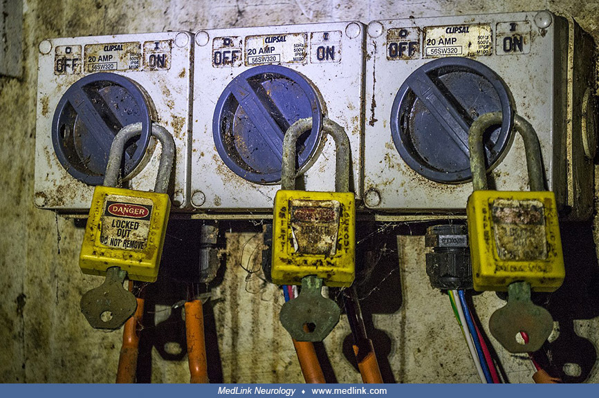
Lockout/tagout (LOTO) procedures involve isolating energy sources and attaching locks and tags to equipment to prevent accidental startup during maintenance or servicing.
By putting a padlock on the source of energy (electrical, hydraulic, or pneumatic pressure, or other energy source), workers may adjust or repair machinery with less likelihood of injury. Lockout scissors are often applied to t...
Examples include using padlocks and hasps on circuit breakers to lock them in the "off" position, preventing accidental energization while electrical work is being performed. In addition, tags are placed on equipment to warn of lockout and to provide further information about the lockout, such as the reason for the lockout, the date, and the name of the authorized employee.
These strategies do not necessarily effectively eliminate the heat hazard from an unintentional electric arc. Consequently, additional protection strategies to reduce occupational exposure are needed. Results of a retrospective study of electrical burn patients admitted to the burn centers in South China indicates that more attention should be paid to preventing electrical burns in male adults, with a particular focus on industrial workers, incidents in the spring and summer, and high-voltage injuries (27).
Neurologic complications of electrical injuries should be differentiated from those due to lightning, particularly in a patient who is not conscious and when history or injury circumstances are not available.
Differentiation from other neurologic conditions resembling complications of electrical injury. The differential diagnosis of the neurologic complications of electrical injuries may include those related to anoxia/hypoxia, cardiac arrest, convulsions, explosion/blast effects, falls, or mechanical trauma from the affected individual being caught by machinery or moving equipment.
Detection of malingering. The rate of malingering is as high as 40% in compensation-seeking workers in general and 25% in electrical injury in particular (52). Many electrical injury patients report neurocognitive impairment or movement disorders. Although entry and exit wounds indicate that the body has absorbed sufficient electricity to cause injury, their presence does not necessarily indicate that the electrical exposure has caused nervous system injury. At the same time, the absence of entry and exit wounds does not necessarily mean that electrical injury has not occurred. Therefore, other indicators of acute CNS dysfunction, such as those used to grade transient brain injury severity (eg, Glasgow Coma Scale score, length of coma or posttraumatic amnesia, structural damage to the nervous system), may also be helpful. Criteria have been developed to provide a systematic, comprehensive, integrated, research-based approach to diagnosing malingered neurocognitive dysfunction (74), and these have been applied to individuals reporting electrical injury (12). Among 11 patients evaluated for neurocognitive impairment after electrical injury, over half of them met the criteria for at least probable malingered neurocognitive dysfunction, and most of the patients with malingered neurocognitive dysfunction lacked evidence of a biologically meaningful exposure to electrical current (12).
|
• Useful diagnostic tools for detecting neurologic sequelae of electrical injury include biomarkers of muscle damage, imaging studies, and electrophysiological studies. |
In general, the diagnostic approach to the neurologic consequences of an electrical injury patient is consistent with the neurologic evaluation of a multi-trauma injury patient. A high degree of clinical suspicion for neurologic involvement is warranted, especially when no or limited information about the responsible electrical events is available and no external wounds indicate localization of exposure.
|
Diagnostic |
Description |
|
Laboratory studies |
Urinalysis, serum biomarkers of muscle damage (eg, creatine kinase), serum myoglobin if urinalysis reveals myoglobinuria. |
|
Radiology studies |
X-rays, CT scans, MRI, MRA, ultrasound, and nuclear medicine examination can evaluate nervous system structural damage and confirm fractures, local tissue edema, focal areas of inflammation, ischemia, thrombosis, or hemorrhage. |
|
Electrophysiological studies |
Electrophysiological studies are guided by the patient’s history and physical examination and may include EKG, electromyography and nerve conduction studies, evoked potential studies, and electroencephalography. Symptoms of peripheral neuropathy should be investigated even when there are minimal cutaneous burns. |
|
• Hospital admission is warranted following exposure to an electrical source of more than 200 V potential, burns involving greater than 15% of body area, and a history of cardiopulmonary resuscitation. | |
|
• Neuropsychiatric sequelae of patients with electrical injuries should be a routine focus of multidisciplinary care. |
American Burn Association Advanced Burn Life Support guidelines outline triage, initial resuscitation, and emergency transport protocols in the emergency or first-response period. Cardiopulmonary resuscitation at the scene can be lifesaving, as was powerfully shown in the "Kiss of Life" Pulitzer Prize-winning photograph by Rocco Morabito in 1967 (75).
On admission to emergency medical services, physical examination, laboratory studies, and diagnostic radiology evaluation are prioritized, depending on the patient’s clinical course. Cardiac monitoring should be initiated in all patients that have experienced anything greater than a minor low-voltage burn.
Neurologic evaluation of the CNS and auditory, visual, and peripheral sensorimotor function can be managed simultaneously by stabilizing cardiorespiratory status and wounds.
Electrical injuries are traditionally divided into low-voltage electric power injuries (less than 1000 V) and high-voltage (greater than 1000 V). In contrast to other types of trauma, high-voltage injuries require a high index of suspicion and awareness of possible manifestations. Electrical injury should be viewed and managed as a multisystem injury (22). Hospital admission is warranted when there is evidence or suspicion of electromechanical contact with an electrical source of more than 200 V potential and capable of more than 200 mA, even without physical findings.
Additional indications for hospital admission include the following occurrences after electrical injury: history of cardiopulmonary resuscitation or cardiac findings, including refractory cardiac arrhythmias; history of loss of consciousness or disorientation; history of fall from a height; associated thermal injury to greater than 15% of the body surface area, or including burn to the hands, feet, face, or groin; suspicion of smoke or vapor inhalation or respiratory distress; spine fractures; serum electrolyte derangements; and compartment syndromes.
Burn care teams must also address the impending neuropsychiatric sequelae of patients with electrical injuries as a routine part of multidisciplinary care. Although the rehabilitation and resuscitation phases of burn care may partly determine neuropsychiatric outcomes, these outcomes may, in turn, determine reconstructive success. For example, posttraumatic stress disorder increases anxiety before, during, and after painful medical procedures. Patients with electrical injuries may benefit from earlier, more focused, and intensive psychiatric support. Although useful guidelines and models exist to systematically address the complex physical morbidities of patients with electrical burns, newer models and strategies are needed to prevent and treat neuropsychiatric sequelae (20).
Limited information is available regarding pregnancy and delivery following electrical shock exposure. Effects of electrical injuries during pregnancy range from a transient unpleasant sensation with no effect on the fetus to sudden maternal and fetal death. Reported adverse effects include fetal heart abnormalities, damage to the fetal CNS, and fetal growth retardation (77).
Considerations regarding anesthesia are related to trauma severity and level of neurologic impairment.
All contributors' financial relationships have been reviewed and mitigated to ensure that this and every other article is free from commercial bias.

Douglas J Lanska MD MS MSPH
Dr. Lanska of the University of Wisconsin School of Medicine and Public Health has no relevant financial relationships to disclose.
See ProfileNearly 3,000 illustrations, including video clips of neurologic disorders.
Every article is reviewed by our esteemed Editorial Board for accuracy and currency.
Full spectrum of neurology in 1,200 comprehensive articles.
Listen to MedLink on the go with Audio versions of each article.
MedLink, LLC
3525 Del Mar Heights Rd, Ste 304
San Diego, CA 92130-2122
Toll Free (U.S. + Canada): 800-452-2400
US Number: +1-619-640-4660
Support: service@medlink.com
Editor: editor@medlink.com
ISSN: 2831-9125
Neurobehavioral & Cognitive Disorders
Jun. 17, 2026
Neuro-Oncology
May. 27, 2026
Neuropharmacology & Neurotherapeutics
May. 14, 2026
Neuro-Oncology
Apr. 30, 2026
Neuropharmacology & Neurotherapeutics
Apr. 23, 2026
Neuropharmacology & Neurotherapeutics
Apr. 20, 2026
Neuro-Ophthalmology & Neuro-Otology
Apr. 07, 2026
General Neurology
Apr. 06, 2026