Neuropharmacology & Neurotherapeutics
Drug-induced neurologic disorders
Dec. 01, 2025
MedLink, LLC
3525 Del Mar Heights Rd, Ste 304
San Diego, CA 92130-2122
Toll Free (U.S. + Canada): 800-452-2400
US Number: +1-619-640-4660
Support: service@medlink.com
Editor: editor@medlink.com
ISSN: 2831-9125
Toll Free (U.S. + Canada): 800-452-2400
US Number: +1-619-640-4660
Support: service@medlink.com
Editor: editor@medlink.com
ISSN: 2831-9125
Nearly 3,000 illustrations, including video clips of neurologic disorders.
Every article is reviewed by our esteemed Editorial Board for accuracy and currency.
Full spectrum of neurology in 1,200 comprehensive articles.
Listen to MedLink on the go with Audio versions of each article.
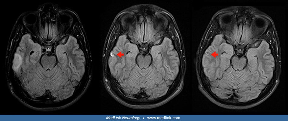
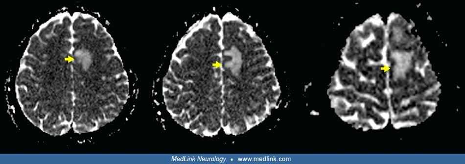
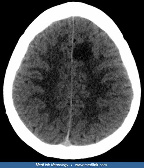
CT scan of the brain in a 31-year-old man working as an underwater fisherman showing a hypodense lesion in the medial left frontal lobe due to decompression sickness. The patient presented with symptoms of paresthesia and difficulty writing. While spearfishing the day before admission, he developed fatigue, headache, and paresthesia, and weakness of the right hemibody. He also reported feeling “confused” and “disoriented” and had difficulty getting out of the water. The patient’s dive profile consisted of a 3-hour dive at a maximum depth of 30 meters, with interimmersion periods of less than 2 minutes on the surface and breath-hold times longer than 2 minutes. Two days earlier he had made another dive of 4 to 5 hours at a similar maximum depth and with a similar dive profile. Neurosonological studies (ie, carotid and vertebral ultrasound and transcranial Doppler studies) of supra-aortic and intracranial arteries showed no atheromatosis or significant hemodynamic alterations. Transthoracic echocardiography was normal, including a negative agitated saline contrast study to assess for a for right-to-left shunt. Total body CT scan showed no evidence of a neoplastic process. (Source: Sánchez-Villalobos JM, Fortuna-Alcaraz ML, Serrano-Velasco L, et al. Breath-hold diving-related decompression sickness with brain involvement: from neuroimaging to pathophysiology. Tomography 2022;8[3]:1172-83. Creative Commons Attribution License [CC BY], https://creativecommons.org/licenses/by/4.0.)