


Neurobehavioral & Cognitive Disorders
Mental status examination
Jun. 17, 2026
MedLink, LLC
3525 Del Mar Heights Rd, Ste 304
San Diego, CA 92130-2122
Toll Free (U.S. + Canada): 800-452-2400
US Number: +1-619-640-4660
Support: service@medlink.com
Editor: editor@medlink.com
ISSN: 2831-9125
Toll Free (U.S. + Canada): 800-452-2400
US Number: +1-619-640-4660
Support: service@medlink.com
Editor: editor@medlink.com
ISSN: 2831-9125
Worddefinition
At vero eos et accusamus et iusto odio dignissimos ducimus qui blanditiis praesentium voluptatum deleniti atque corrupti quos dolores et quas.
Divers who have experienced pressures greater than two atmospheres absolute may develop decompression sickness if they ascend too rapidly. Decompression sickness may be mild, with only limb and joint pain ("bends," type I), or serious, with neurologic, cardiac, and pulmonary manifestations (type II). Divers with a patent foramen ovale are more likely to develop severe forms of decompression sickness than divers without a right-to-left shunt. Treatment in a pressure chamber is essential for recovery, and detailed decompression tables are used to prevent and treat decompression sickness.
|
• Decompression sickness usually occurs during rapid ascent from depth after diving but may also occur in rapid ascent to high altitudes from sea level. | |
|
• Systemic manifestations may involve the nervous system. | |
|
• Decompression sickness can be avoided by gradual ascent, but if decompression sickness occurs, it is treated by hyperbaric recompression. | |
|
• Hyperbaric oxygen is useful in treating decompression sickness with neurologic manifestations. |
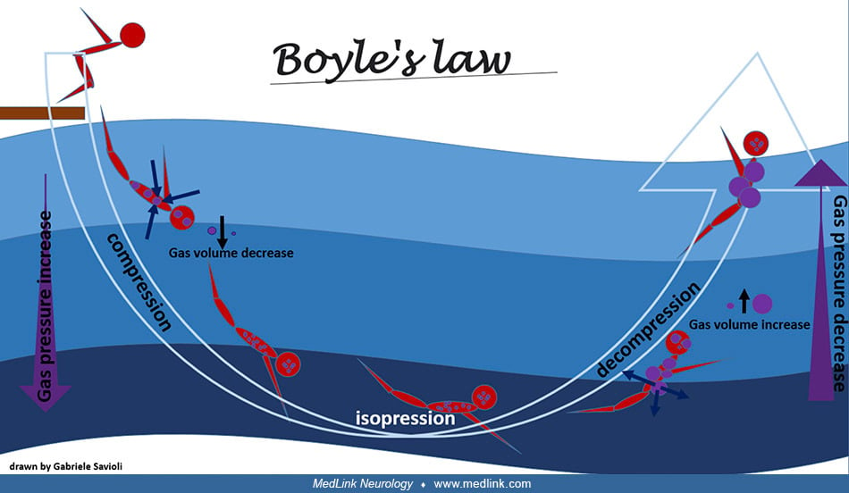
Boyle's law. In 1660, Anglo-Irish natural philosopher Robert Boyle (1627-1691) published the first controlled experiments with "rarified air," obtained by reducing the pressure of the air (20; 21; 140).
Boyle found by 1662 that (in modern language) for a fixed mass of an ideal gas kept at a fixed temperature, pressure, and volume are inversely proportional (21). For his experiments, Boyle relied on an air pump devised by English polymath Robert Hooke (1635-1703).
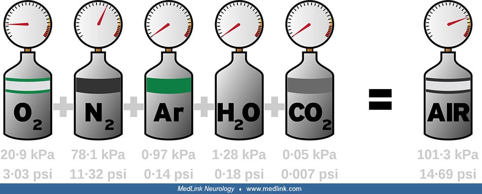
Dalton's law of partial pressures. In a mixture of gases, as in air, each constituent gas has a partial pressure that is the pressure of that constituent gas as if it alone occupied the entire volume of the original mixture at the same temperature. This is Dalton's law, named after English chemist and physicist John Dalton (1766-1844), who determined this experimentally in 1802 (35).
At sea level, where atmospheric pressure is 760 mm Hg, the percent of the total composition and the partial pressures of the various gases are approximately as follows: 78.6%, 597 mm Hg for nitrogen; 20.9%, 159 mm Hg for oxygen; 0.04%, 3.0 mm Hg for water; and 0.004%, 0.3 mm Hg for carbon dioxide. For comparison, the percent of the total composition and partial pressures of alveolar air are as follows: nitrogen 74.9%, 569 mm Hg; oxygen 13.7%, 104 mm Hg; and water 6.2%, 40 mm Hg.
Henry’s law. Henry’s law, formulated by English chemist William Henry (1774-1836) in 1803, states that, at a constant temperature, the solubility of a gas is directly proportional to the pressure that the gas exerts on the solution (63).
When equilibrium is reached, the solution is described as "saturated," but if the pressure is then reduced, the tissues become effectively supersaturated, and the gases leave the solution and may form gas bubbles. Due to the metabolic activity of oxygen and carbon dioxide, and the comparatively marked inactivity of nitrogen, it is nitrogen that is by far the most problematic. To determine the time necessary for the clearance of supersaturated nitrogen without biological damage, decompression tables have been generated based on biomathematical models (but relying on the experience and fitness of Navy divers). Even following protocols established for safe decompression, problems may occur (eg, interindividual variation, flying in a commercial aircraft within 12 to 18 hours after diving, etc.), which has led to the appreciation that safety factors must be built into the estimated "safe" dive times.
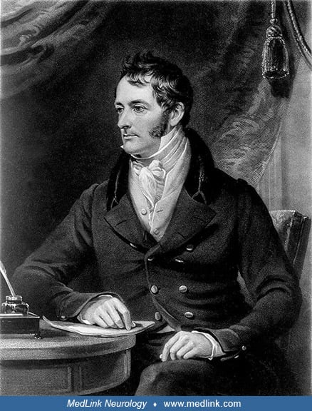
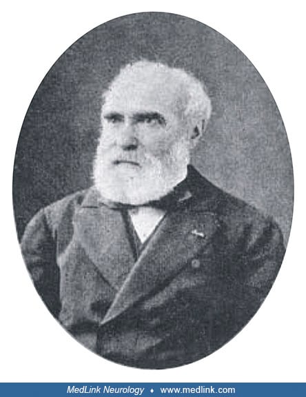
The Bert and Smith "effects" of oxygen toxicity. In 1878, French zoologist, physiologist, and politician Paul Bert (1833-1886) was the first to determine the acute toxicity of high oxygen concentrations in "La Pression Barometrique” (13; 14). Bert was a student of French physiologist Claude Bernard (1813-1878).
Among the students is Paul Bert (back row, third from left). (From a photogravure of the picture of 1889 by Leon L'Hermite in the Sorbonne. Creative Commons 4.0 International License [CC BY 4.0], https://creativecommons.org/lic...
Bert applied an apparatus of French physiologist Denis Jourdanet (1815-1892) that was intended for the therapeutic use of compressed or "expanded" air. Bert experimented on himself with "superoxygenated air," that is, air with an increased partial pressure of oxygen.
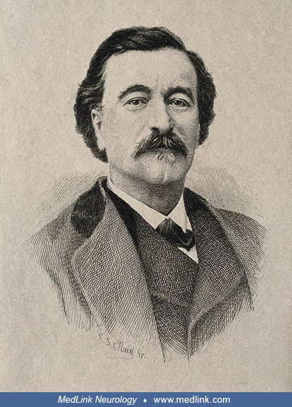
In 1878, Bert demonstrated convulsions in larks exposed to air at 15 to 20 atmospheres absolute, and the neurotoxic effects of oxygen at increased pressure were subsequently called the "Bert effect" (13). Then in 1899, Scottish pathologist and physiologist J(ames) Lorain Smith (1862-1931), while trying to reproduce the "Bert effect," noticed fatal pneumonia in rats after 4 days of exposure to 73% oxygen at one atmosphere absolute, which marked the discovery of pulmonary toxicity of oxygen at increased partial pressure--the "Smith Effect" (117; 57; 61).
Smith discovered pulmonary toxicity of oxygen at increased partial pressure, the "Smith effect" (Smith JL. The pathological effects due to increase of oxygen tension in the air breathed. J Physiol 1899;24[1]:19-35). (Source: Pr...
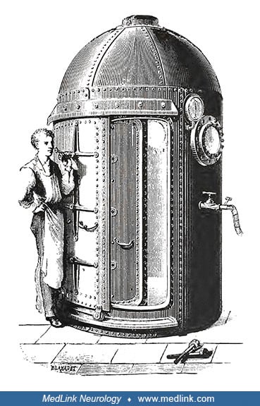
Development of an underwater breathing apparatus. English wool merchant John Lethbridge (1675-1759) invented the first underwater diving machine in 1715 to facilitate the salvage of shipwrecks.
English astronomer, mathematician, and physicist Edmond Halley (1656-1742), later famous for predicting the return of the comet named in his honor, developed a practical diving bell in 1717.
Halley's diving bell was constructed of wood, covered in lead, and weighted to keep it correctly oriented underwater. It was 8 feet high, 5 feet in diameter at the bottom, and 3 feet in diameter at the top. Fresh air was supplied by two lead-lined barrels with bung holes at the bottom. The barrels were alternately lowered to the sea floor, where an attendant pulled the tube up toward the bell, allowing air to be forced by pressure from the barrel and into the bell. Halley and four others were able to remain on the sea floor at a depth of 9 to 10 fathoms (54 to 60 feet) for 90 minutes. An improved version of Halley's diving bell was developed by Edinburgh confectioner and amateur engineer Charles Spalding (1738-1783) in 1775, and a more sophisticated underwater breathing apparatus was developed by Kleingert in 1798.
An improved version of Halley's diving bell by Edinburgh confectioner and amateur engineer in 1775. (Source: Hill L. Caisson sickness and the physiology of work in compressed air. London: Edward Arnold, 1912. Public domain.)
In 1819, German-born British engineer Augustus Siebe (1788-1872) invented a diving helmet for an "open dress" form of diving to provide greater mobility to the diver.
This consisted of a metal helmet and shoulder plate attached to a water-tight jacket. The helmet was fitted to an inlet valve to which a flexible air-supply tube was attached. The tube was connected to an air pump, and the force of the pressurized air kept the water from rising in the jacket.
In 1878, pioneering English diving engineer Henry Albert Fleuss (1851-1933) was granted a patent for an apparatus that improved rebreathers, ie, a breathing apparatus that absorbs the carbon dioxide of a user's exhaled breath to permit the rebreathing (recycling) of the unused oxygen and inert gas content (45).
It consisted of a rubber mask connected to a breathing bag, with 50% to 60% oxygen supplied from a copper tank and carbon dioxide scrubbed using a rope yarn soaked in a strongly alkaline solution of caustic potash (potassium hydroxide), the system giving a working duration of about 3 hours. In 1879, Fleuss demonstrated the utility of his device by submerging himself in a water tank for an hour and then, 1 week later, by diving to a depth of 5.5 meters in open water. Fleuss's apparatus was first used under operational conditions in November 1880 by Alexander Lambert (c1837 to 1892), the lead diver of the Severn Tunnel construction project to build a railway tunnel linking South Gloucestershire in the west of England to Monmouthshire in south Wales under the estuary of the River Severn. After being trained by Fleuss, Lambert was able to close a submerged sluice door in the tunnel that had foiled hard-hat divers due to the strong water currents and the danger of their air supply hoses becoming fouled on submerged debris. The same apparatus was later used several times to rescue mine workers.
American environmental medicine and diving medicine specialist Christian James Lambertsen (1917-2011) was principally responsible for developing the rebreathers used by U.S. Navy frogmen for underwater warfare during World War II.
Lambertsen designed a series of rebreathers in 1940 and in 1944, first calling his invention simply "breathing apparatus." Consequently, the U.S. Navy considers Lambertsen to be "the father of the Frogmen." Later, after the war, Lambertsen called his invention LARU (an acronym for Lambertsen Amphibious Respiratory Unit). In 1952, he again changed his invention's name to SCUBA (Self-Contained Underwater Breathing Apparatus).
Diving regulator technology was subsequently invented by French engineer Émile Gagnan (1900-1984) and French naval officer and oceanographer Jacques-Yves Cousteau (1910-1997) in 1943.
Although the Gagnan-Cousteau invention was unrelated to rebreathers and came after Lambertson's apparatus, Lambertson's "SCUBA" term is now generally applied to the Gagnan-Cousteau invention.
Use of different breathing mixtures. English-born American engineer and inventor Elihu Thomson (1853-1937) is best known for his electrical innovations and entrepreneurism (eg, in 1892 his Thomson-Houston Electric Company merged with the Edison General Electric Company to become the General Electric Company), but he was also instrumental in the development of different breathing mixtures for diving and caisson work (29).
As early as 1873, Thomson published a paper on the inhalation of nitrous oxide, nitrogen, hydrogen, and other gases and gaseous mixtures (129), and in 1927, he specifically proposed the use of helium in deep diving and caisson work (130; 131).
Dysbarism. Dysbarism is a general term that encompasses disturbances in the human body resulting from a change in atmospheric pressure. Dysbarism encompasses five subentities: decompression sickness, barotrauma, gas embolism, inert gas narcosis, and oxygen toxicity.
Decompression sickness. Decompression sickness is one of several forms of dysbarism, ie, disturbances in the human body resulting from a change in atmospheric pressure. Divers, miners, tunnel workers, and caisson workers will experience decompression sickness if they progress too quickly to a lower environmental pressure. Rapid ascent to high altitudes in an aircraft with an uncompressed cabin can also produce decompression sickness. The first and least severe symptoms are characterized by limb and joint pain. In more severe cases, with ascent from greater depths, after longer bottom times, or with more rapid ascent or decompression, other nervous system, cardiac, or pulmonary symptoms may occur.
The condition was well known among caisson workers, and it was, therefore, called "caisson disease." Other terms used to describe the condition are "the bends" (limb and joint pain), "the chokes," and "hits."
|
• Dysbarism encompasses five subentities: decompression sickness, barotrauma, gas embolism, inert gas narcosis, and oxygen toxicity. | |
|
• Neurologic manifestations of decompression sickness while diving appear during or immediately after ascent. | |
|
• A common neurologic manifestation of decompression sickness is myelopathy. | |
|
• Livedo racemosa (often incorrectly labeled "cutis marmorata"), due to gas emboli in the cutaneous circulation, precedes neurologic symptoms and almost always indicates a right-to-left shunt. |
Dysbarism. Dysbarism is a general term that encompasses various pathologies that result when the body experiences an increase or decrease in atmospheric pressure at a rate or duration that exceeds the capacity of the body to safely adapt (111). Although the most common cause of dysbarism is underwater diving, it can also occur with aviation and space exploration as well as compressed air tunnel and caisson work ("caisson's disease" --a caisson is a large watertight chamber, open at the bottom, from which the water is kept out by air pressure and in which construction work may be carried out underwater) (132; 53).
Dysbarism encompasses five subentities: decompression sickness, barotrauma, gas embolism, inert gas narcosis, and oxygen toxicity.
Decompression sickness. Symptoms of decompression sickness develop rapidly and appear during or immediately after ascent. They usually occur within 3 hours of surfacing but may sometimes take as long as 24 to 36 hours to manifest. They are mainly seen in individuals who have experienced pressures greater than two atmospheres absolute.
In a retrospective study from 2011 to 2023 in the neurology department of a military hospital in Tunis, 10 subjects exhibited cerebral involvement, all male professional divers with a mean age of 41 years (88). Symptoms manifested within 10 minutes of surfacing in 66% of cases and included sensory-motor deficits, vertigo, and headache.
Aviation decompression sickness can also occur, for example, with rapid decompression of cabin pressures (89), or with military parachutist dispatchers exposed repeatedly to high altitude (25,000 feet) (33). However, events that occur at flight altitudes below 17,000 feet (5182 meters) or with rapid decompression pressure changes under 0.3 atm, decompression sickness is less likely to be the etiology of the presenting symptoms.
Golding and colleagues proposed a dichotomous classification of decompression sickness: a minor form (Type I) affecting the musculoskeletal system and characterized by limb and joint pain ("the bends"), and a major form (Type II) involving the neurologic ("hits"), pulmonary ("the chokes"), and cardiac systems (53). Neurologic manifestations are present in 10% to 15% of decompression sickness cases. Isolated cutaneous manifestations (ie, livedo racemosa) are generally considered to be type I decompression sickness. Circulator shock is considered to be type II decompression sickness.
Neurologic manifestations of decompression sickness. A common neurologic manifestation of decompression sickness is myelopathy (137; 97; 94; 135; 38). Symptoms of spinal cord involvement usually appear early, typically within 3 minutes of surfacing. They are characterized by leg weakness with walking difficulties, leg numbness and paresthesia, and bladder dysfunction. Other reported symptoms include intense nausea, localized back pain, penile erection, and fecal incontinence (38). The severity varies from mild sensory symptoms to paraplegia or even tetraplegia. In most cases, the segmental level of the responsible lesion is around the sixth to eighth thoracic segment, which is the area most susceptible to ischemia. Decompression sickness may, however, involve any part of the spinal cord. Fortunately, in many cases of spinal decompression sickness, the symptoms gradually disappear, leaving the patient asymptomatic, although slight reflex abnormalities may persist as evidence of earlier damage.
Spinal cord decompression sickness presenting as Brown-Sequard syndrome, with MRI abnormalities corresponding to infarction in the territory of a posterior spinal artery, may improve considerably following hyperbaric oxygen therapy (97; 135); the diagnosis is mainly based on clinical findings rather than MRI (135). Gas bubbles may also block the anterior spinal artery, causing weakness in both lower extremities (137).
Some divers develop decompression sickness with acute cerebral hemispheric dysfunction, manifesting variously as hemiparesis, aphasia, hemianopsia, Anton syndrome (ie, visual anosognosia due to cortical blindness) (08), memory loss, convulsions, and coma.
Inner ear decompression sickness may manifest as vertigo and hearing loss, but so may inner ear barotrauma (85; 93; 112). Development of post-dive benign paroxysmal positional vertigo may occur due to nitrogen bubble formation within the semicircular ducts (36; 37). In an observational case series of 13 divers presenting acutely with inner ear decompression sickness in Plymouth, UK, between July 2021 and January 2024, average test values for vertical perception, posturography, dynamic gait index, and patient-reported outcome measures improved by discharge and at the 3-month follow-up despite 67% showing a persisting positive head impulse test or nystagmus in the dark on video nystagmography (125).
Livedo racemosa (often mislabeled "cutis marmorata"). Cutis marmorata, livedo reticularis, and livedo racemosa have been improperly considered synonyms when, in fact, they refer to phenomena with different appearances and pathophysiology (60). Cutis marmorata is the most commonly used term in the context of decompression sickness and is a form of livedo reticularis; however, this is "a physiological and benign, livid circular discoloration with a net-like, symmetric, reversible, and uniform pattern" (60).
Cutis marmorata is a physiologic form of livedo reticularis observed commonly on the legs on exposure to cold, with gradual resolution on rewarming; it occurs primarily in cold children and slim young women (eg, after being in cool water) (60). It is a physiological vasospastic response to coldness or systemic disease with a consequent decline of tissue oxygen saturation, particularly in the periphery of the vascular distribution of small cutaneous arterioles (60). The characteristic symmetric and uniform cyanotic mottling pattern is related to the vascular anatomy of normal skin. The microanatomical structure of the cutaneous blood supply is arranged in a series of 1 to 3 cm cones at the margins of which the zone of arterial predominance wanes, while the superficial venous plexus becomes more prominent.
Therefore, physiologic or pathologic processes, like livedo reticularis, that impede cutaneous blood flow may produce a regular, net-like coloration pattern of closed rings in the predominantly venous areas at the margins of the cones.
Skin pattern of livedo reticularis (top and bottom left) and urticaria (bottom right) for comparison with livedo racemosa. (Source: Hartig F, Reider N, Sojer M, et al. Right/left shunt. Front Physiol 2020;11:994. Copyright © 20...
In contrast, decompression-associated skin discolorations correspond to the "pathological, irregular, broken netlike pattern" of livedo racemosa, a pathologic and painful disorder typically found on the limbs, trunk, and buttocks, which intensifies with higher ambient temperature (60). Livedo racemosa is caused by thrombotic or embolic vascular occlusion of arterioles with resultant local perfusion deficits and hypo-oxygenation, causing an irregular reddish-blue or violaceous net-like mottling of the skin.
The pattern of livedo racemosa is caused by vascular occlusion of arterioles with irregular, local perfusion deficiency leading to a reddish blue, irregular mottling of the skin. The cases shown in this figure were not related ...
With decompression sickness, the embolic obstructions of the ascending or feeding arterioles consist of gas bubbles, whereas in other settings, the emboli may consist of fibrin, cholesterol, or calcium phosphate.
Several hypotheses have been proposed to explain livedo racemosa (52; 80; 47): (1) local formation of bubbles in cutaneous blood vessels; (2) arterialization of venous bubbles across a right-to-left shunt augmented by local amplification of bubble size on reaching supersaturated skin via the arterial circulation; and (3) arterialized venous gas bubbles embolizing the brainstem, where the autonomic nervous system regulates skin blood vessel dilation and constriction (52; 80; 24). Of these, the most likely is that livedo racemosa results from arterialized venous bubbles that pass to the cutaneous circulation via a right-to-left shunt (hypothesis 2), possibly with bubble augmentation on reaching supersaturated skin.
Livedo racemosa is generally caused by thrombotic or embolic occlusion of cutaneous arteries, which suggests that decompression-associated livedo racemosa is caused by arterial gas emboli (hypothesis 2) (60). Moreover, when proper techniques are used, investigators have uniformly identified significant right-to-left shunts in patients with decompression-associated livedo racemosa (82; 47; 60; 09). In one study of four divers with livedo racemosa (albeit mislabeled cutis marmorata), all four had the following: (1) large right-to-left shunts, (2) numerous small bubbles visibly moving within the skin microvasculature, and (3) no bubbles visible in adjacent areas of normal skin (47). In another case of livedo racemosa (also mislabeled cutis marmorata), the presence of intravascular air was confirmed by CT (77). These findings discount hypotheses 1 and 3 and confirm the validity of hypothesis 2.
Decompression sickness-related circulatory shock. A patient who presented with neurologic symptoms and hypovolemic shock after two risky dives had multiple cerebral and pulmonary thromboembolisms on MRI (84).
Another recreational diver dove to a depth of 218 feet for 43 minutes while breathing air and developed nitrogen narcosis. He ascended from depth slowly according to the staged decompression recommendations of his dive computer, but ultimately he exhausted his air supply and was forced to surface, having omitted 6.5 hours of required air decompression (06). After requesting emergency medical services, he lost consciousness on the surface and was rescued from the water; he was then intubated and mechanically ventilated. The clinical presentation was remarkable for loss of consciousness, hypotension, livedo racemosa, peripheral edema, and severe hypoxia requiring mechanical ventilation. Shock worsened during hyperbaric oxygen therapy, requiring aggressive volume resuscitation with crystalloid plus albumin in addition to multiple vasopressors. After 37 hours of hospitalization, blood pressure normalized, and vasopressors were tapered and discontinued. He ultimately required 10 days of mechanical ventilation and was discharged on day 21 with mild decompression sickness-related neurologic deficits.
Early hyperbaric oxygen therapy can reduce the neurologic deficits associated with decompression sickness plus shock, and cardiopulmonary and renal functions can normalize with aggressive volume expansion and pressors to counteract refractory hypotension (84; 06).
Decompression sickness-related damage to other organs. Rarely, decompression sickness can present with other organ damage, such as severe acute kidney injury (28).
Barotrauma. Barotrauma is physical tissue damage caused by a pressure difference, usually between an unvented space inside the body and surrounding gas or fluid, but it can occur superficially as well; for example, mask squeeze barotrauma results from failure to equalize the air space created between the face mask and the face.
The damage is due to shear or overstretching of tissues. As a gas-filled space expands or contracts, it can cause damage to the local tissue. This is a phenomenon well-known to deep-sea fishermen who pull up deep-dwelling fish that have experienced barotrauma by rapid ascent in the nets.
A fish brought up from a considerable depth with a distended swim bladder prolapsed, everted, and projecting from its mouth (after Regnard). (Source: Hill L. Caisson sickness and the physiology of work in compressed air. London...
Pulmonary compression barotrauma may occur during very deep descents during breath-hold diving. As lung volume decreases to or below residual volume, mucosal edema, vascular engorgement, pulmonary edema, and hemorrhage may occur as nonventilated areas fill with liquid to counteract the negative pressure. Affected individuals may experience chest pain, dyspnea, and hemoptysis on ascent.
Ear barotrauma may affect the external, middle, and inner ear. This may occur on descent (compression) or ascent (decompression), depending on the circumstances that cause pressures to be unbalanced across the tympanic membrane or the round and oval windows. For example, if the external auditory canal is blocked by cerumen, the higher air pressure from the tank regulator passes through the Eustachian tube, causing the affected tympanic membrane to bulge outward and possibly to rupture with resultant severe pain and bloody otorrhea. If, instead, the Eustachian tube is blocked, and the diver is unable to "clear" the passage and balance the pressures with a Valsalva maneuver, middle ear pressure will be less than the pressure of the water against the tympanic membrane, causing the affected tympanic membrane to bulge inward and possibly to rupture with (again) resultant severe pain and bloody otorrhea. Less commonly, the round or oval window may rupture with Eustachian tube dysfunction, causing severe, acute vertigo, a potentially life-threatening situation at depth.
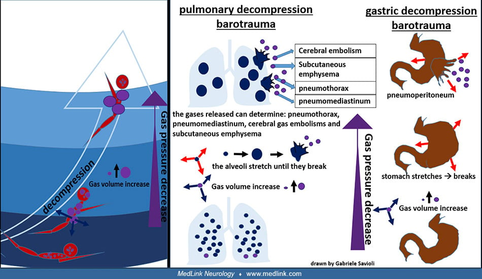
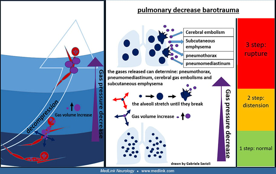
Decompression barotraumas can damage the lungs and gastrointestinal tract. As the pressure decreases with ascent, the volume occupied by the gases increases (Boyle’s law), and the excess gas (ie, excess for the available volume of the organ that contains it) is exhaled from the lungs and expands within the gastrointestinal tract to be expelled, if possible, from the mouth or anus. If the surplus is retained, the tissues that contain the gas are stretched and damaged. Pulmonary decompression barotraumas may cause cerebral arterial gas embolism, subcutaneous emphysema, pneumothorax, and pneumomediastinum. Gastrointestinal decompression barotraumas may cause gastrointestinal rupture and pneumoperitoneum.
Pulmonary barotrauma is a significant reason (15%) for emergency calls to the Divers Alert Network (DAN) involving diving minors, likely because of psychological immaturity, suboptimal management of adverse situations, and inadequate supervision (62).
Sinus barotrauma may result from blocked or intermittently blocked sinuses. The inability to maintain a balance between the air pressure within the sinuses and the ambient pressure (which is matched by the regulator for the inspired air or gas mixture) causes the pressure inside the sinuses to become lower than the ambient pressure during descent; if some high-pressure gas enters the sinus during the dive and partially compensates for the higher ambient pressure at depth, a reverse situation may occur on ascent. The pressure differences alone can cause intense sinus pain, vascular engorgement, and epistaxis. In addition, during ascent, compression of the maxillary branch of the trigeminal nerve in the maxillary sinuses can cause infraorbital paresthesia that usually resolves within several hours without treatment.
Gas embolism. Gas embolism can occur when a pressure gradient allows air to enter the bloodstream or when bubbles form within the bloodstream. The bubbles then obstruct blood flow in distal vessels. Gas emboli may complicate barotrauma, decompression sickness, and mixed forms. Arterial gas embolism is usually precipitated by rapid ascent, breath holding, or preexisting lung disease.
Inert gas narcosis. Inert gas narcosis (also known as nitrogen narcosis, "rapture of the deep," the "Martini effect," and the "narcs") is a reversible alteration in consciousness that occurs while diving at depth (83). It is caused by the anesthetic effect of certain inert gases at high pressure. Narcosis produces a state similar to drunkenness (alcohol intoxication), so the relation of depth to narcosis has been referred to informally as "Martini's law.” Inert gas narcosis results in the feeling of one martini for every 10 meters (33 ft) below 20 meters (66 ft) depth, so the effect starts at 30 meters or 100 feet, with a pressure of approximately four atmospheres absolute.
Narcosis may be completely reversed in a few minutes by ascending to a shallower depth, with no long-term effects. Nitrogen narcosis rarely develops into a serious problem if the divers are aware of its symptoms and ascend to manage it. Diving much beyond 40 meters (130 feet) is outside the scope of recreational diving. Special training in the use of various helium-containing gas mixtures (eg, heliox or trimix) is required to dive at greater depths for military or commercial purposes and minimizes the risks of narcosis and oxygen toxicity. These mixtures prevent narcosis by replacing some or all of the nitrogen fraction of the breathing gas with non-narcotic helium.
Manifestations of inert gas narcosis may include: (1) feelings of tranquility, exhilaration, giddiness, anxiety, depression, or paranoia; (2) impaired concentration and multi-tasking ability; (3) impaired judgment and decision-making ability; (4) increased reaction time and impaired coordination; (5) vertigo and visual or auditory disturbances; and (6) reduced perception of cold discomfort and impaired shivering (with a consequent faster decline in core body temperature, aggravating many of the problems of inert gas narcosis) (58; 66).
Oxygen toxicity. Oxygen toxicity (or oxygen poisoning) results from breathing oxygen at higher-than-normal partial pressures (34). In particular, breathing oxygen at partial pressures above 1.4 atmospheres absolute (ie, a partial pressure of more than approximately 220 mm Hg) can produce acute neurotoxicity. Activities and situations associated with increased risk of oxygen toxicity include underwater diving breathing air, hyperbaric oxygen therapy, exposure to prolonged high oxygen levels, and prematurity. Acute oxygen toxicity typically manifests with CNS effects ("the Bert effect"), whereas chronic toxicity presents with mainly pulmonary effects ("the Smith effect"). CNS effects can include fasciculations and myoclonus, nausea, tinnitus, dysphoria, and generalized convulsions; pulmonary effects can include pleuritic chest pain, substernal heaviness, coughing, dyspnea, damage to the pulmonary epithelium, pulmonary (intra-alveolar) edema, inactivation of surfactant, interstitial thickening and fibrosis, and atelectasis.
Deep diving. Diving at depths of more than 50 meters of seawater is designated deep diving. Breathing air is not advised at those depths due to the risk of nitrogen narcosis, a toxic effect of the high partial pressure of nitrogen. Therefore, other gas mixtures are used (eg, heliox, a mixture of helium and oxygen, or triox, a mixture of oxygen, helium, and nitrogen).
Diving with heliox to depths greater than 150 meters produces signs and symptoms of high-pressure neurologic syndrome. The syndrome includes tremor in the upper extremities, impaired memory, dizziness, nausea, and in severe cases, myoclonic jerks and even unconsciousness. High-pressure neurologic syndrome in a dive chamber is accompanied by EEG changes with increased theta (73).
Deep diving requires a prolonged period of compression and a decompression period that may extend over several days. Even after uncomplicated deep dives, transitory focal neurologic changes have been reported, although it is unclear whether these represent an unmasking of otherwise asymptomatic lesions from prior decompression sickness or are instead evidence of new lesions.
Long-term neurologic sequelae of decompression sickness are common among professional offshore divers (126). Among 208 retired Norwegian offshore divers, 163 (78%) reported episodes of decompression sickness, with neurologic decompression sickness in 41 (20%) (126). Divers with a history of neurologic decompression sickness had significantly more neurologic findings on tests of motility, coordination, and sensation (126).
Long-term follow-up of 30 divers treated with hyperbaric oxygen for decompression sickness in 2009 and 2010 revealed that a quarter had long-term residual symptoms (128). More severe presentations of decompression sickness are more likely to have severe long-term sequelae (eg, gait disorders and sphincter incontinence) (18). In decompression sickness with myelopathy, the improvement in MRI findings is not necessarily associated with improved clinical status. Initial motor impairment, further aggravation during transfer to the hyperbaric facility, and development of sphincter dysfunction are indicators of poor prognosis, regardless of the treatment.
In a population-based study, those with decompression sickness had a 3.8-fold increased risk of developing psychiatric disorders and a 5.7-fold increased risk of developing sleep disorders (134).
In a study of 59 military divers with decompression sickness, the main independent risk factor for poor outcome (ie, sensory and motor deficits or bladder dysfunction) was the severity of neurologic manifestations at onset, and recovery was not significantly improved by prompt administration of recompression treatment (17).
In a prospective study of 28 divers with neurologic decompression sickness at a hyperbaric facility, those who presented more than 17 hours after surfacing were likely to have more intense symptoms than those who presented earlier (101). However, delayed initiation of hyperbaric oxygen therapy was not associated with a need for more hyperbaric treatment or worse outcomes.
The therapeutic response in patients with inner ear decompression sickness remains poor. Treatment with 100% oxygen followed by recompression in a hyperbaric chamber will improve the condition of most patients, but two thirds have incomplete recovery (51). Time to recompression does not clearly influence clinical outcome. Vestibular symptoms are more prevalent in retired offshore divers than in age-matched controls, presumably because of prior decompression sickness: among retired divers, 28% reported dizziness, 14% had vertigo, and 25% had an unsteady gait (54).
Dysbaric osteonecrosis (aseptic bone necrosis) most commonly involves the femoral head but may involve other joints, including the humeral head (25). Symptoms may appear months after diving (25). It is an occupational hazard for commercial and navy divers, but it is also seen in sport divers.
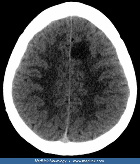
A 31-year-old man working as an underwater fisherman presented with a 1-day history of paresthesia and difficulty writing (110). While spearfishing, he developed fatigue and headache as well as paresthesia and weakness of the right hemibody. He also reported feeling “confused” and “disoriented” and had difficulty getting out of the water. The patient’s dive profile consisted of a 3-hour dive at a maximum depth of 30 meters, with inter-immersion periods of less than 2 minutes on the surface and breath-hold times longer than 2 minutes. Two days earlier, he had made another dive of 4 to 5 hours at a similar maximum depth and with a similar dive profile. CT scan of the brain showed a hypodense lesion in the medial left frontal lobe due to decompression sickness.
CT scan of the brain in a 31-year-old man working as an underwater fisherman showing a hypodense lesion in the medial left frontal lobe due to decompression sickness. The patient presented with symptoms of paresthesia and diffi...
Neurosonological studies (ie, carotid and vertebral ultrasound and transcranial Doppler studies) of supra-aortic and intracranial arteries showed no atheromatosis or significant hemodynamic alterations. Transthoracic echocardiography was normal, including a negative agitated saline contrast study to assess for a right-to-left shunt. Total-body CT scan showed no evidence of a neoplastic process.
Given the patient's history of breath-hold diving and the clinical and neuroimaging findings, the patient was transferred to a hyperbaric chamber. Recompression was carried out in the hyperbaric chamber at 2.8 atmospheres absolute. With a progressive resolution of symptoms, treatment was completed using United States Navy Table 6. Total elapsed time is 285 minutes, ascent time between stops (18 to 9 meters and 9 to 0 meters) is 30 minutes (3 meters per 10 minutes), and maximum pressure is 18 meters of seawater.
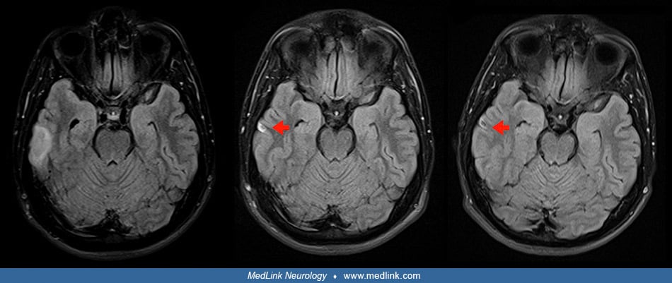
MRI studies done 4 and 5 days after clinical onset showed a left frontal cortico-subcortical hyperintense lesion on FLAIR images due to decompression sickness. Axial diffusion-weighted imaging (DWI b1000) sequences did not show high-signal-intensity lesions.
FLAIR images 4 and 5 days after onset due to decompression sickness. MRI done 4 days after clinical onset (left and center) and 5 days after clinical onset (right) show a left frontal cortico-subcortical hyperintense lesion (re...
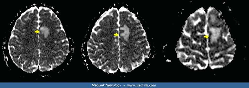
Axial apparent diffusion coefficient maps showed a hyperintense lesion in the left frontal lobe. Axial gadolinium-enhanced 3D T1-weighted imaging (Gd T1WI) showed the presence of different areas of enhancement with irregular morphology in the left frontal area, not suggestive of tumor enhancement. Over a 4-month period, the left frontal lesion gradually evolved with lessening hyperintensity on axial FLAIR images, ultimately leaving a small area of encephalomalacia.
Axial apparent diffusion coefficient maps show a hyperintense lesion in the left frontal lobe (yellow arrows). MRI done 4 days after clinical onset (left and center) and 5 days after clinical onset (right). (Source: Sánchez-Vil...
Axial gadolinium-enhanced 3D T1-weighted imaging (Gd T1WI) shows the presence of different areas of enhancement with irregular morphology in the left frontal area (blue arrow), not suggestive of tumor enhancement. MRI done 4 da...
Axial FLAIR images initially (left), at 1 month (center), and 4 months (right). By 1 month, the lesion is minimally hyperintense (yellow arrow). By 4 months, there is a small area of encephalomalacia (yellow arrow). (Source: Sá...
MRI studies done 4 and 5 days after clinical onset also showed a right temporal cortico-subcortical hyperintense lesion with slight mass effect due to decompression sickness. Over a 4-month period, the right temporal lesion gradually evolved with lessening hyperintensity on axial FLAIR images, ultimately leaving a small area of encephalomalacia.
MRI done 4 days after clinical onset (left, left center, and right center columns) and 5 days after clinical onset (right) column. Axial fluid-attenuated inversion recovery (FLAIR) images (left column) show a right temporal cor...
Axial FLAIR images initially (left), at 1 month (center) and 4 months (right). By 1 month the lesion is minimally hyperintense (red arrow). By 4 months, there is a small area of encephalomalacia (red arrow). (Source: Sánchez-Vi...
|
• Breathing air or gas mixtures under increased pressure leads to increased nitrogen saturation of tissues. | |
|
• On decompression (eg, on ascent from diving or with rapid gain in altitude during flying), gas bubbles may form in the blood and embolize to the CNS and other organs. | |
|
• Embolic lesions in the CNS are facilitated by a right-to-left shunt (eg, patent foramen ovale). |
Decompression sickness. Decompression sickness is caused by the formation of gas bubbles in the blood and tissues during or after a decrease in environmental pressure (decompression).
As ambient pressure increases, the inspired pressure of the inert gas (most often diatomic nitrogen, N2) is greater than its arterial partial pressure, which causes the tissues to absorb the gas until the two pressures are balanced. A rapid decrease in pressure (eg, with ascent while diving) causes the dissolved nitrogen to return to its gas form while it is still in the blood or tissues, creating intravascular "bubbles."
These bubbles can cause mechanical obstruction of a vein or an artery (ie, venous or arterial gas embolism) and can also produce an inflammatory response with activation of complement, coagulation factors, and platelets.
Ascending and descending degeneration is shown in the sections above and below these levels (Boycott and Damant). (Source: Hill L. Caisson sickness and the physiology of work in compressed air. London: Edward Arnold, 1912. Publ...
This can lead to the formation of thrombi, an increase in vascular permeability and interstitial edema, and stasis of the microcirculation with tissue hypoperfusion and ischemia. Divers, aviators, and astronauts are at risk of decompression sickness when ambient pressure reductions exceed a critical threshold.
Gas bubbles are present in venous blood after ascent from water depths as shallow as 3 meters. Free venous gas does not invariably lead to decompression sickness because most bubbles are filtered out by the pulmonary capillaries and do not enter the arterial circulation. However, the foramen ovale remains open, or patent, in 25% to 34% of the adult population, with detectable shunting in 8% to 10% (64), and divers with a patent foramen ovale have a significantly increased risk of developing decompression sickness (22; 118); the risk increases with increasing grade of the right-to-left shunt (118). Large right-to-left shunts due to a patent foramen ovale are associated with an increased risk of spinal cord decompression sickness with thoracolumbar involvement (49). In addition, filtration is size-dependent, as small bubbles may escape entrapment. Small emboli do not necessarily cause infarction but may disturb the blood-brain barrier, inducing what has been termed the "perivenous syndrome" (74).
There is a high prevalence of patent foramen ovale in divers suffering from inner ear decompression sickness manifested by vestibular and cochlear symptoms following dives while breathing helium-based mixtures (56).
In a few reported autopsies of apparently unaffected divers, CNS degeneration and vasculopathy have resembled the abnormalities found after decompression sickness. Subclinical damage associated with small emboli is common, especially in the midbrain, as demonstrated by MRI (74).
Predisposing factors for decompression sickness. Risk factors for decompression sickness include strenuous activity during the dive, dehydration, and dive depths exceeding 30 meters of seawater (127). Exercise-induced muscle damage from eccentric work that is characterized by reduced strength and delayed-onset muscle soreness leads to increased formation of venous gas emboli during subsequent hypobaric exposure (ie, acute decompression) (55). Persons with right-to-left shunt (eg, patent foramen ovale) have an increased risk of developing neurologic complications even after recreational scuba diving. Whether cigarette smoking increases the risk of developing decompression sickness is unclear, but it has been debated.
Yo-yo diving (ie, a series of short-duration dives alternating with similar periods on the surface) was previously considered to reduce bubble formation and risk of decompression sickness based on studies in rats. Subsequently, concern was raised that yo-yo divers may have an increased risk of neurologic decompression sickness, which has been supported by detection of gas bubbles in the left ventricle and neuronal injury in the spinal cord in a study using pigs (102).
A patent foramen ovale or other left-to-right shunt patent foramen ovale can lower the threshold for venous gas embolism tolerance with a tendency for earlier and more abundant arterialization during decompression (105; 106).
An experimental study in rats has shown that pretreatment with sildenafil, a phosphodiesterase-5 blocker used for the treatment of erectile dysfunction, promotes the onset and severity of neurologic decompression sickness, possibly related to vasodilation with increased cerebral blood flow (16).
Decompression sickness-related circulatory shock. Decompression sickness-related shock results from bubble-endothelial interactions, which cause a transient capillary leak syndrome associated with plasma extravasation, hemoconcentration, and hypovolemia (06).
Altitude-related decompression sickness. Cases of altitude-related decompression sickness, some with CNS manifestations, were reported in an Air Force training facility during pressure reduction in a chamber to simulate altitudes between 25,000 and 35,000 feet (7,500 and 10,668 meters) (23). The number of altitude-related decompression sickness incidents with CNS manifestations among United States Air Force U-2 pilots in the period from 2002 to 2009 was greater than experienced by United States military aviators in the preceding 47 years, due in large part to longer, more frequent high-altitude exposure (75). The U-2 can climb to 60,000 feet in less than 45 minutes, which far exceeds the altitude and rate of ascent of a passenger airliner, which might take 30 to 40 minutes to reach a cruising altitude of only 35,000 feet. A review of decompression sickness cases associated with the NASA altitude physiological training program at Johnson Space Center indicated an incidence of 1.16 cases per 1000 exposures with significant heterogeneity across studies (32); denitrogenation time, exposure pressure, and exposure time were associated with the probability of decompression sickness.
Because decompression sickness is one of the most likely adverse effects among pilots of supersonic, high-altitude aircraft, they may require hyperbaric oxygen treatment before resuming active duty (109). An F/A-18D pilot, who presented with neurologic symptoms following a loss of cabin pressure at altitude, required multiple hyperbaric oxygen therapy sessions over several days due to the recurrence of symptoms on resumption of flying.
Neurologic decompression sickness in 50 high-altitude pilots was associated with a significant increase in the volume of hyperintense white matter lesions on MRI, especially in the insula; this was attributed to hypobaric exposure rather than hypoxia because all pilots were maintained on 100% oxygen throughout their flights (100).
Among United States military personnel exposed to extreme altitude-chamber exposures (from 29 tests comprising 708 altitude chamber exposures), important contributors to hypobaric decompression sickness included decompression "dose" (based on higher tissue ratio or bubble growth index), male gender, and high-intensity exercise at altitude (31).
Most cases of altitude-related decompression sickness occur among individuals exposed to altitudes of 25,000 feet or higher (105; 106).
Barotrauma. Barotrauma occurs when gas pressures across a membrane or surface differ sufficiently to cause damage.
Greater than 90% of the human body is either water or bone, which are effectively incompressible, leaving those structures that are filled with air or gas to be most directly affected by pressure changes: the middle ear, the Eustachian tube, the sinuses, the lungs, and the gastrointestinal tract.
Barotrauma can occur either with an increase (eg, on descent while diving) or with a decrease (eg, on ascent while diving) of the ambient pressure. Under Boyle's Law, described by Anglo-Irish natural philosopher Robert Boyle (1627-1691), as external pressure increases, the gas volume in the body cavities containing air or other gas decreases (eg, lungs, middle ear, sinuses, gastrointestinal tract, etc.), as it will also in a face mask in which the pressure is not equalized with the ambient pressure. The opposite occurs when external pressure decreases. Iatrogenic barotrauma may, of course, also occur during intubation and mechanical ventilation (ie, “ventilator-associated lung injury” or “ventilator-induced lung injury”).
Inert gas narcosis. The mechanism of inert gas narcosis is not well understood, but it may result from gas dissolving into nerve membranes and causing a temporary disruption in nerve transmissions (83). Although the effect was first observed with air (due to the nitrogen content), other gases, including argon, krypton, and hydrogen, cause very similar effects at higher than atmospheric pressure. Although some of these effects may be due to antagonism at NMDA receptors and potentiation of GABAA receptors, this is not likely to be the result of chemical bonding to receptors because these manifestations can be produced by breathing the very chemically inactive gas argon at high pressure.
|
Pressure (bar) |
Depth (m) |
Depth (ft) |
Symptoms |
|
1 to 2 |
0 to 10 |
0 to 33 |
Minor or none |
|
2 to 4 |
10 to 30 |
33 to 100 |
Mildly impaired reasoning |
|
4 to 6 |
30 to 50 |
100 to 165 |
Sense of well-being |
|
6 to 8 |
50 to 70 |
165 to 230 |
Sleepiness |
|
8 to 10 |
70 to 90 |
230 to 300 |
Stupefaction |
|
10+ |
90+ |
300+ |
Euphoria, mania, or depressive states |
|
Note: a bar is defined as 100,000 Pa (100 kPa) or slightly less than the current average atmospheric pressure on Earth at sea level (1 atm, approx. 1.013 bar). Within a few percent, 1 bar = 100 kPa, approx. 1 at (or atmospheres absolute), approx. 1 atm, approx. 750 Torr, approx. 14.5 psi. | |||
Oxygen toxicity. Oxygen toxicity is caused by oxygen-derived free radicals generated by mitochondrial oxidoreductive processes, certain extra-mitochondrial enzymes (eg, xanthine/urate oxidase), auto-oxidative reactions, and phagocytes during bacterial killing. These free radicals create lipid peroxidations, especially in cell membranes, and disrupt nucleic acids and protein synthesis, leading to membrane rupture and cell death.
The incidence of decompression sickness in the general diving population is relatively low, ranging from 0.01% to 0.095% depending on the diving environment and type of diving performed (138).
Between 2002 and 2006, the incidence of decompression illness in Australia was 10.74 per 100,000 dives or lower, and the diving-associated mortality rate among scuba divers was 0.57 per 100,000 dives (96).
A retrospective study of the incidence and characteristics of decompression sickness in Denmark from 1999 to 2013 identified 205 cases, a more than 10-fold increase from 1966 to 1980 (128). The most frequent symptoms in this series were paresthesia (50%), pain (42%), and vertigo (40%). In the subgroup of divers treated in 2009 and 2010, a quarter had long-term residual symptoms.
Among 55 experienced and highly trained Finnish technical divers, 27 episodes of decompression sickness occurred in 2983 dives, for an incidence of 91 episodes of decompression sickness per 10,000 technical dives (136). All reported decompression illness symptoms were mild, and only one diver received hyperbaric oxygen therapy, whereas 21% used first-aid oxygen. Eventually, all divers recovered without residual symptoms.
Risk factors for decompression sickness include (1) long, deep dives; (2) use of aggressive decompression protocols; (3) omitted decompression; (4) rapid ascent; (5) strenuous work at depth; (6) cold on decompression; and (7) repetitive dives (especially over multiple days) (64).
A high-grade patent foramen ovale is a risk factor for unprovoked decompression sickness in recreational divers (71). The foramen ovale remains open, or patent, in 25% to 34% of the adult population, with detectable shunting in 8% to 10% (64). In relatively small cohorts of divers with a known patent foramen ovale, the incidence of decompression sickness ranges from 0.5% to 1.8% (133; 95).
Medical personnel in hyperbaric treatment centers have a significantly increased occupational risk for decompression sickness while attending patients inside the multiplace hyperbaric chamber (87).
Decompression sickness is a pervasive problem among diver fishermen ("artisanal fisheries"), particularly in some underdeveloped countries, such as the Dominican Republic (44; 99; 115), and also occurs in commercial breath-hold divers (eg, Ama of Japan) (86; 39; 19).
|
• To prevent the formation of bubbles leading to decompression sickness, divers should limit their ascent rate to 10 meters (33 feet) per minute. | |
|
• Known risk factors (eg, heavy exercise and alcohol prior to a dive) should be avoided. | |
|
• Divers should avoid flying within 24 hours after their last dive. |
Diving-related decompression sickness can usually be prevented when the dive profile is conservative (eg, avoiding decompression dives), when the frequency of dives is limited, when dive limits (time and depth) are not exceeded, and when care is taken during controlled ascent to normal pressure. The United States Navy and commercial diving companies have published detailed decompression tables. To prevent the formation of bubbles leading to decompression sickness, divers should limit their ascent rate to 10 meters (33 feet) per minute. In addition, predetermined "decompression stops" may be necessary for specified periods at specified depths, depending on the gas mixture and bottom time.
There is some theoretical justification for the use of helium-containing trimix over air for advanced recreational diving to decrease the inflammatory response associated with gas emboli and thereby minimize the risk and severity of decompression sickness. However, in physically fit, advanced recreational divers, no difference in inflammatory markers was identified in divers using either trimix or air when using a similar diving profile of identical duration (108).
Although exercise several hours before diving is beneficial, heavy exercise just before or after diving may increase the risk of decompression sickness (98). Cold exposure, recent alcohol use, and dehydration increase risk and should be avoided. General fitness should also be encouraged, with weight loss and tobacco cessation counseling provided as necessary.
Pre-dive hydration with 30% of the recommended daily water intake before scuba diving effectively suppresses the formation of bubbles after diving and decreases the risk of decompression sickness (59).
Divers should avoid flying within 24 hours after their last dive, and empiric data from a study of 316 divers suggest that most (79%) observed this recommendation; only a small proportion (1.9%) boarded a flight with a pre-flight surface interval of less than 12 hours (119). Diagnosed and treated decompression sickness developed during and after flight in four divers with pre-flight surface intervals of at least 24 hours (1.6%) compared with double that frequency for divers with pre-flight surface intervals of less than 24 hours (3.1%). Surprisingly, 15 divers (4.8%) boarded a plane with perceived symptoms of decompression sickness.
Pre-dive conditioning measures, such as endurance exercise in a warm environment, oral hydration, and normobaric oxygen breathing, can reduce the risk of decompression sickness in scuba divers (48).
Hyperoxia facilitates gas exchange across the bubble surfaces and increases the share of bubble content contributed by oxygen rather than inert gas (eg, nitrogen) (02; 03). Pre-breathing high concentrations of oxygen before ascent can decrease the frequency of decompression sickness (eg, pre-breathing with 100% oxygen before and during the flight in high-altitude military aircrews and using oxygen-enriched gas mixes in commercial and military diving) (75; 105; 106). Hyperoxic excursions in an altitude chamber from 24,000 feet (7315 meters) to 18,000 feet (5486 meters) reduced, but did not fully prevent, the occurrence of venous gas emboli (02; 03; 2022). Pre-breathing was not clearly beneficial for short-duration hypobaric exposures (26).
Another approach for the prevention of decompression sickness is whole-body vibration preconditioning, or the use of a "mini trampoline," both of which reduce the formation and delay the manifestations of diving-related or high-altitude-induced venous gas emboli (43; 90).
The diagnosis of decompression sickness is usually straightforward when the symptoms appear during diving operations or rapid decompression. It should be remembered, however, that symptoms occurring during diving are not necessarily related to variations in the environmental pressure, as the stress of diving may exacerbate pre-existing chronic diseases (eg, chest pain due to heart disease, dyspnea due to chronic lung disease).
The symptoms of neurologic decompression sickness may resemble those of thromboembolic cerebrovascular disease, but decompression sickness more commonly affects the spinal cord. However, symptoms attributed to a stroke immediately after a scuba dive should not deter a trial of hyperbaric oxygen therapy, even when there are other risk factors for stroke (eg, age and atrial fibrillation) (123). Delays in recognizing decompression sickness and initiating treatment may delay and preclude full recovery (123).
Pulmonary barotrauma can lead to arterial embolization of gas bubbles, which can mimic decompression sickness, particularly if cerebral or spinal symptoms occur within minutes of surfacing from a dive. This can happen, for example, (1) in panicked ascent without expiring (even with very short and shallow dives with compressed gas) (92); (2) with explosive decompression accidents (in aircraft or dive chambers); or (3) with pulmonary blebs or bullae during gradual, controlled ascents especially if there is airway narrowing or focal stenosis (eg, by mucous plugs).
Inner ear barotrauma and inner ear decompression sickness both manifest with cochleovestibular symptoms (vertigo plus hearing loss), causing difficulties in differential diagnosis and possibly delaying (or leading to inappropriate) treatment (36; 37; 85; 93; 112; 120). Extended lifetimes of bubbles at hyperbaric pressure may contribute to inner ear decompression sickness during saturation diving (41). Development of post-dive benign paroxysmal positional vertigo in conjunction with decompression sickness may occur due to nitrogen bubble formation within the semicircular ducts (36; 37). Hemorrhage into the semicircular ducts or canals may also occur (65).
Alternobaric vertigo may also cause diagnostic confusion but is a benign condition caused by a temporary incongruency in middle ear pressures due to incomplete or insufficient equalization, typically while ascending (ie, moving from higher pressure to lower pressure) (112; 46).
Cases of factitious decompression sickness have been reported (81).
High-pressure neurologic syndrome, which can occur in deep-sea diving beyond a depth of 100 meters, is characterized by tremor, cognitive disturbances, and characteristic EEG changes. Symptoms generally clear rapidly on ascending.
Divers with a patent foramen ovale are more liable to develop decompression sickness, as air bubbles can pass directly from the venous to arterial blood.
Pulmonary barotrauma may occur in divers because an increase in the volume of gas entrapped in the lungs during ascent leads to alveolar rupture, entry of the gas into systemic circulation via the pulmonary veins, and systemic air embolism. The gas may track around the vessels, leading to mediastinal emphysema. Rupture of peripheral alveoli may lead to pneumothorax. The pulmonary changes are accompanied by hypoxemia, pulmonary hypertension, and respiratory distress. Bubbles trapped in the lungs may also cross the pulmonary circulation due to a reduction in their size on compression.
Dysbaric bone necrosis (dysbaric osteonecrosis, aseptic bone necrosis) has been reported after a single hyperbaric air exposure with inadequate decompression. It usually results from gas bubbles entering end arteries in the bone and is most common in compressed air workers.
|
• The diagnosis of decompression sickness is based primarily on history and clinical features rather than diagnostic studies. | |
|
• If a person has been diving to less than 10 meters or has been exposed to pressures lower than two atmospheres absolute, the symptoms are unlikely to be caused by decompression sickness. | |
|
• Primary screening for patent foramen ovale is not routinely indicated in divers or aircrews. | |
|
• A causative patent foramen ovale is strongly suggested by any of the following: (1) the onset of decompression sickness symptoms immediately after a physical isometric effort or Valsalva-like maneuver, (2) the onset of decompression sickness symptoms after low-risk flights (eg, at low cabin altitudes or at high altitudes but for a short time), or (3) dives that are close to the limits of “no-decompression diving” or close to the required mandatory decompression stops. | |
|
• A physical isometric effort or Valsalva-like maneuver immediately preceding the onset of symptoms is highly suggestive of a causative patent foramen ovale. | |
|
• Echocardiography is indicated in patients who have experienced a decompression sickness event, as they may have a right-to-left shunt that predisposes them to further episodes of decompression sickness (among other adverse outcomes). |
Decompression sickness. The diagnosis of decompression sickness is based primarily on history and clinical features and, in particular, should consider the characteristics of the exposure, symptoms and signs, and comorbid conditions. If a person has been diving to less than 10 meters or has been exposed to pressures lower than two atmospheres absolute, the symptoms are unlikely to be caused by decompression sickness (other than with decompression sickness associated with rapid ascent to high altitude).
Although rare, hypoalbuminemia at initial presentation due to capillary leak accurately predicts the neurologic manifestations of decompression sickness in scuba divers (50).
When neurologic decompression sickness is suspected, MRI studies of the brain and spinal cord should be performed. MRI revealed acute subcortical and hyperintense white matter lesions in U-2 pilots with neurologic decompression sickness (76); as is the case with diving decompression sickness, however, imaging played no role in acute diagnosis but was considered vital for determining fitness for return to flying.
In a study of 10 professional divers with cerebral decompression sickness, MRI revealed diverse patterns, such as corpus callosum hyperintensities on T2 FLAIR in five cases, an acute ischemic stroke in one patient, and punctiform or nodular lesions in others (88). DWI abnormalities suggested both cytotoxic and vasogenic edema.
Suspected barotrauma. A chest x-ray and high-resolution CT of the chest should be done to detect pneumothorax or mediastinal air, especially if there are manifestations of pulmonary barotrauma. High-resolution CT may demonstrate gas in the peritoneal cavity or extrapulmonary thorax and within joints (78). An untreated pneumothorax is an absolute contraindication for hyperbaric treatment due to the possibility of converting it to a tension pneumothorax as intrapleural air expands on decompression.
Right-to-left shunts. Primary screening for patent foramen ovale is not routinely indicated in divers or aircrews (105; 106). A recreational diver with an incidental patent foramen ovale should be counseled according to the context, size of shunt, and the individual’s preferences and expected compliance (105; 106). Primary patent foramen ovale screening can be suggested to professional divers with non-modifiable high-risk characteristics for decompression sickness (105; 106). Military pilots assigned to frequent and prolonged flight activity at less than 280 mmHg barometric pressure or more than 25,000 feet can undergo patent foramen ovale screening.
A causative patent foramen ovale is strongly suggested by any of the following: (1) the onset of decompression sickness symptoms immediately after a physical isometric effort or Valsalva-like maneuver, (2) the onset of decompression sickness symptoms after low-risk flights (eg, at low cabin altitudes or at high altitudes but for a short time), or (3) dives that are close to the limits of “no-decompression diving” or close to the required mandatory decompression stops (105; 106). In cases of decompression sickness during low-risk activities, patent foramen ovale screening should be performed (105; 106). The possibility of simultaneous or alternative intrapulmonary shunts should always be considered because patent foramen ovale closure will reduce the risk of paradoxical venous gas emboli but will have no effect on the development of venous gas emboli or pulmonary shunts (105; 106).
A physical isometric effort or Valsalva-like maneuver immediately preceding the onset of symptoms is highly suggestive of a causative patent foramen ovale (105; 106). The same applies if symptoms occur after low-risk flights, such as those at low cabin altitudes or at high altitudes but for a short time, or dives such as those that are close to the limits of “no-decompression diving” or close to the required mandatory decompression stops, according to the utilized decompression model.
Echocardiography is indicated in patients who have experienced a decompression sickness event, as they may have a right-to-left shunt that predisposes them to further episodes of decompression sickness (among other adverse outcomes) (09). Transthoracic echocardiography with antecubital injection of agitated saline bubbles using proper technique is often revealing (104). However, intra-femoral injection of agitated saline bubbles may be necessary to counteract the hemodynamic effects of a large Eustachian valve (a ridge-like remnant of a fetal structure in the inferior right atrium that directed incoming oxygenated blood to the foramen ovale, also known as the "valve of the inferior vena cava") (09).
Ultrasonic monitoring of bubbles during decompression is not of demonstrated benefit for predicting neurologic sequelae of decompression sickness. However, detection of marrow bubbles with MRI after musculoskeletal decompression sickness can predict subsequent dysbaric osteonecrosis (124). Dysbaric bone necrosis can be detected on x-rays, but often after a significant delay.
Other studies. 99mTc HMPAO SPECT images of decompression sickness reveal areas of reduced perfusion, which may be subclinical but nevertheless persist in most cases (122; 114; 40). However, the clinical significance of perfusion deficits found in divers remains unclear (67; 121).
Forensic studies of diving deaths. To determine the cause of diving-related deaths, forensic pathologists employ accident reconstruction in conjunction with analysis of diving monitoring data, witness reports, toxicology studies, postmortem CT examination, and gas analysis (location and quantity) in the body of the corpse (141).
On-site first aid. First aid of decompression sickness consists of on-site 100% oxygen, immobilization (ie, no unnecessary movement), and fluid administration; telephone consultation with a diving medicine specialist is recommended (79). Unfortunately, on-site first aid administration of oxygen is frequently omitted (27).
Medical transport. Rapid movement of a patient with decompression sickness may present difficulties when the site of the hyperbaric treatment facility is a considerable distance away. Fortunately, the movement of patients with decompression sickness by low-level helicopter flight is both safe and effective, especially when a pressurized aircraft is neither available nor practical. In one study, patients were transferred by low-level helicopter flight without evident complications when the helicopter stayed within 200 feet (61 meters) above ground level of the take-off point, but symptoms of decompression sickness did worsen when this altitude was exceeded (107). A more recent study found that subjective symptoms or oxygen levels of patients with decompression sickness may improve when the patient is transported in physician-staffed emergency helicopter flights less than 300 meters in height with the administration of oxygen and fluids (103).
Recompression and hyperbaric oxygen. The treatment of decompression sickness is, first and foremost, recompression. Treatment aims to reduce bubble growth, promote gas clearance, and counteract ischemia and hypoxia in the affected tissues. Therefore, if possible, treatment should be initiated before pathologic changes become irreversible.
Treatment is performed in a hyperbaric chamber following various hyperbaric oxygen protocols (11; 12; 05; 10; 72).
U.S. Navy Treatment Table 6 has been widely used for mild decompression sickness, but shorter treatment schedules may be better, requiring fewer treatments until symptom resolution and more often accomplishing complete symptom resolution after the initial treatment (10). Hyperbaric oxygen therapy is effective in restoring and preserving neurologic function in the “perivenous syndrome” associated with decompression sickness in both animal and clinical studies (74).
In a retrospective study, hyperbaric oxygen therapy administered within 24 hours of the onset of type 1 decompression sickness was associated with rapid relief of symptoms after a single treatment (91). Seasoned military divers showed a faster response after recompression with fewer residual symptoms than commercial or recreational divers.
A patient with high-altitude decompression sickness recovered completely following hyperbaric oxygen therapy, recompression, and extracorporeal oxygenation (116). In another case, an unconscious stowaway was discovered in a wheel well of a Boeing 747-400F but recovered completely with hyperbaric oxygen therapy (113).
Despite oxygen first aid being given infrequently and long delays before definitive treatment, treatment outcome following decompression and hyperbaric oxygen therapy is generally satisfactory; at the end of hyperbaric oxygen treatment, most divers (65%) recover completely, but many have significant residua (27).
In-water recompression. In-water recompression involves placing the affected diver back in the water at a depth of 9 meters with a prolonged pure oxygen-breathing period followed by gradual ascent to the surface (44; 139). In-water recompression has been used by several navies and in remote areas where local recompression chambers are not available. In-water recompression is not without risk and should only be carried out by divers with suitable equipment and practical training.
Decompression sickness-related circulatory shock. The pathophysiology and typical clinical course of decompression sickness-related shock support the use of aggressive but time-limited volume expansion and pressors to counteract hypovolemia (06). Hyperbaric oxygen therapy should be strongly considered for decompression sickness even in the face of extreme critical illness with refractory shock and respiratory failure (06).
Drug therapy. No drugs have been clearly demonstrated to benefit human decompression sickness. Aspirin is used frequently for the treatment of decompression sickness, but its efficacy has not been established by controlled studies. Fluoxetine, an antidepressant with anti-inflammatory properties, decreases the incidence of experimental decompression sickness in mice (15). Statin-mediated lipid reduction may reduce bubble generation via alterations in plasma rheology and surface tension. The use of high-dose steroids in decompression sickness is controversial.
Patent foramen ovale management. In divers with a history of major neurologic decompression symptoms, transesophageal echocardiography should be performed to exclude a right-to-left shunt (eg, patent foramen ovale, atrial septal defect, etc.). Divers with a history of decompression sickness and a cardiac right-to-left shunt should stop diving until the shunt has been corrected. Patent foramen ovale closure may reduce the risk of decompression sickness among divers, but adverse events may occur in up to 8% of those undergoing surgical correction (68; 42; 01). No difference is observed in the occurrence of venous bubbles between patent foramen ovale and patent foramen ovale closure groups, but catheter-based patent foramen ovale closure eliminates arterial bubbles after simulated dives (70). Divers with a history of decompression sickness may resume diving after patent foramen ovale closure with reasonable safety, but a conservative diving profile is recommended when there is a residual shunt after patent foramen ovale closure to minimize the risk of recurrent decompression sickness events (42). The subgroup of divers with an unclosed patent foramen ovale and a previous episode of serious decompression sickness may not be safe to dive, even within conservative limits (22).
In a meta-analysis of four observational studies with a total of 309 divers (141 with PFO closure and 168 without PFO closure), patent foramen ovale closure was associated with a significantly lower incidence of decompression sickness (PFO closure 2.8% versus no closure 11%; relative risk 0.29; 95% CI: 0.10 to 0.89; number needed to treat for an additional beneficial outcome = 11) (01). Adverse events occurred in 7.6% of patent foramen ovale closures, including tachyarrhythmias and bleeding.
In a study of a graded-strategy approach, divers with a high-grade patent foramen ovale were offered either catheter-based patent foramen ovale closure (the closure group) or advised conservative diving (high grades), whereas divers with a low-grade shunt were advised conservative diving (low grades), and those with no patent foramen ovale continued unrestricted diving (controls) (69). Among 702 divers who continued diving, decompression sickness incidence decreased significantly in all groups except the controls. During follow-up, there were no decompression sickness events in the closure group; decompression sickness incidence was similar to the controls in the low-grade group but remained higher in the high-grade group.
Information on pregnancy outcomes in humans is limited, with inconsistent data on diving and birth defects, spontaneous abortions, and stillbirth. The safest choice during pregnancy is to avoid diving (30). The fetus is not protected from decompression sickness and is at risk of congenital malformations and gas embolism. However, hyperbaric oxygen therapy can be considered if a pregnant woman develops decompression sickness. Such treatments have been carried out safely in pregnant women for other indications and are considered safe for the fetus in the later months of pregnancy, although experimental evidence raises the possibility of congenital malformations in the early stages of pregnancy.
All contributors' financial relationships have been reviewed and mitigated to ensure that this and every other article is free from commercial bias.

Douglas J Lanska MD MS MSPH
Dr. Lanska of the University of Wisconsin School of Medicine and Public Health has no relevant financial relationships to disclose.
See ProfileNearly 3,000 illustrations, including video clips of neurologic disorders.
Every article is reviewed by our esteemed Editorial Board for accuracy and currency.
Full spectrum of neurology in 1,200 comprehensive articles.
Listen to MedLink on the go with Audio versions of each article.
MedLink, LLC
3525 Del Mar Heights Rd, Ste 304
San Diego, CA 92130-2122
Toll Free (U.S. + Canada): 800-452-2400
US Number: +1-619-640-4660
Support: service@medlink.com
Editor: editor@medlink.com
ISSN: 2831-9125
Neurobehavioral & Cognitive Disorders
Jun. 17, 2026
Neuro-Oncology
May. 27, 2026
Neuropharmacology & Neurotherapeutics
May. 14, 2026
General Neurology
May. 13, 2026
Neuro-Oncology
Apr. 30, 2026
Neuropharmacology & Neurotherapeutics
Apr. 23, 2026
Neuropharmacology & Neurotherapeutics
Apr. 20, 2026
Neuro-Ophthalmology & Neuro-Otology
Apr. 07, 2026