Stroke & Vascular Disorders
Subarachnoid hemorrhage
Jun. 26, 2025
MedLink, LLC
3525 Del Mar Heights Rd, Ste 304
San Diego, CA 92130-2122
Toll Free (U.S. + Canada): 800-452-2400
US Number: +1-619-640-4660
Support: service@medlink.com
Editor: editor@medlink.com
ISSN: 2831-9125
Toll Free (U.S. + Canada): 800-452-2400
US Number: +1-619-640-4660
Support: service@medlink.com
Editor: editor@medlink.com
ISSN: 2831-9125
Nearly 3,000 illustrations, including video clips of neurologic disorders.
Every article is reviewed by our esteemed Editorial Board for accuracy and currency.
Full spectrum of neurology in 1,200 comprehensive articles.
Listen to MedLink on the go with Audio versions of each article.
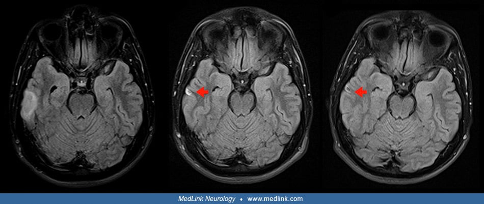
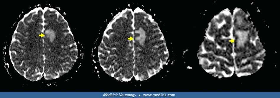
MRI done 4 days after clinical onset (left, left center, and right center columns) and 5 days after clinical onset (right) column. Axial fluid-attenuated inversion recovery (FLAIR) images (left column) show a right temporal cortico-subcortical hyperintense lesion with slight mass effect (red arrows). Axial diffusion-weighted imaging (DWI) sequences (left center column) did not show a high-signal intensity of lesions, whereas axial apparent diffusion coefficient maps (right center column), show a hyperintense lesion in this location (yellow arrows). (D,H) Axial gadolinium-enhanced 3D T1- weighted (Gd T1WI) images (right column) show the presence of a slight enhancement of irregular morphology (blue arrow). (Source: Sánchez-Villalobos JM, Fortuna-Alcaraz ML, Serrano-Velasco L, et al. Breath-hold diving-related decompression sickness with brain involvement: from neuroimaging to pathophysiology. Tomography 2022;8[3]:1172-83. Creative Commons Attribution License [CC BY], https://creativecommons.org/licenses/by/4.0.)