Introduction
Overview
This article describes the neurologic effects of various biological agents that could be used in biowarfare and bioterrorism. Clinical manifestations and pathophysiology of the important agents from a neurologic perspective (particularly anthrax and botulism) are described.
Key points
|
• Anthrax has been used as a biowarfare and bioterrorism agent, and repeated (though unsuccessful) attempts have been made to use botulinum toxin as a bioterrorism agent. |
|
• The Biological Weapons Convention, entered into force in 1975, prohibits the development, production, acquisition, transfer, stockpiling, and use of biological and toxin weapons. |
|
• Severe hemorrhagic meningitis regularly occurs in anthrax and is often accompanied by severe systemic disease, most likely transmitted by spore inhalation. |
|
• Neurologists are most likely to become involved in primarily diagnosing bioterrorist attacks that use botulinum toxin through oral ingestion or inhalation. |
|
• Characteristics that make a pathogen a high risk for bioterrorism include a low infective dose, ability to be aerosolized, high contagiousness, and hardiness (ie, survival in various environmental conditions). |
|
• In the CDC 3-tier categorization of bioterrorism agents and diseases, Category A is comprised of high-priority agents that pose a risk to national security because (1) they can be easily disseminated or transmitted from person to person, (2) they have the potential for major public health impact (eg, high case-fatality rates), (3) they will likely cause public panic and social disruption, and (4) they require special measures for public health preparedness. |
|
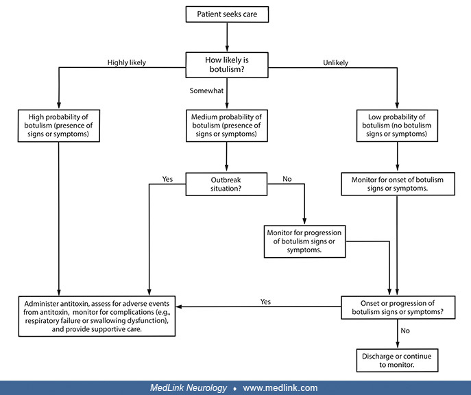
• Diagnostic and management recommendations for anthrax meningitis and botulism vary across conventional (noncrisis), contingency, and crisis settings. |
Historical note and terminology
Biological weapons and bioterrorism agents include replicating agents (eg, bacteria, spores, viruses) and nonreplicating agents (eg, toxins).
Individuals in many cultures have employed biological agents for execution, assassination, and murder. However, many of the claims concerning the use of biological weapons in ancient history are not supported by rigorous evidence and, although seemingly cited in the introductions to numerous articles, editorials, and monographs on bioweapons and bioterrorism, are unreferenced and uncritical rehashing of speculative and dubious claims, often reiterated and amplified on social media (50). Supported and plausible claims include reported attempts to poison water supplies with diseased carcasses (eg, the hurling of plague- or smallpox-infested material or bodies across protective barriers) and deliberate attempts to spread smallpox in enemy armies or populations using biologically inoculated fabrics and people.
The Geneva Protocol (1925): "no use" versus "no first use" of biological weapons. The Geneva Protocol (formally known as the Protocol for the Prohibition of the Use in War of Asphyxiating, Poisonous or other Gases, and of Bacteriological Methods of Warfare) was signed in Geneva in June 1925 and entered into force in February 1928. The Geneva Protocol prohibits the use of biological weapons, a first important milestone towards a comprehensive ban on biological weapons. Several States ratified the Protocol with reservations concerning the Protocol’s applicability and regarding the use of chemical or biological weapons in retaliation, effectively rendering the Geneva Protocol a "no-first-use agreement."
Bioterrorism and "neuroterrorism." Bioterrorism is defined as the intentional use of biological, chemical, nuclear, or radiological agents to cause disease, death, or environmental damage. Bioterrorism that intentionally targets the nervous system has been labeled “neuroterrorism” (19).
Because civilian panic and disruption of institutional operations are prominent intentions (and outcomes) of terrorist attacks, panic and psychologically determined “me-too” symptomatology will contribute significantly to the diagnostic and management burden (53).
History of the United States Biological Warfare Program. The United States did not begin an offensive biological warfare program until 1941, when concern about the German and Japanese biological warfare threats motivated the United States to begin biological weapon development (122).
During World War II, Allied intelligence services suspected the German military might use botulinum toxin as a biological weapon, especially during preparations for Operation Overlord, the Allied invasion to liberate Europe (122). However, despite these persistent concerns, botulinum toxin was not part of the German military arsenal (even if some German scientists had tried to use French pre-war military research for this purpose). The misinformation spread by British and American intelligence services stimulated military biological weapons research at the Porton Down facilities in England and at Camp Detrick in the United States.
During the next 28 years, the United States initiative evolved into a military-driven research and acquisition program shrouded in controversy and secrecy. Most research and development was done at Fort Detrick, Maryland, whereas production and testing occurred at Pine Bluff, Arkansas, and Dugway Proving Ground in Utah. Field testing was done secretly with simulants and actual agents disseminated over wide areas.
Initially, a small defensive effort paralleled the weapons development and production program. Later, the program became entirely defensive with the decision of President Richard Nixon in 1969 to halt offensive biological weapons production and the subsequent agreement in 1972 at the international Biological Weapons Convention never to develop, produce, stockpile, or retain biological agents or toxins. The U.S. Biological Defense Research Program now conducts research to develop physical and medical countermeasures to protect service members and civilians from the threat of biological warfare.
Prior "simulated germ-warfare attack" by the U.S. military: Operation Sea-Spray (1950). Operation Sea-Spray was a secret U.S. Navy biological warfare experiment conducted in September 1950. In the experiment, Serratia marcescens and Bacillus globigii bacteria were sprayed over the San Francisco Bay Area in California to assess the vulnerability of a city like San Francisco to a bioweapon attack (40; 118). From September 20th until September 27th, the Navy used giant hoses to spray a fog of bacteria (species then believed to be harmless) from a ship cruising off the shore of San Francisco. Based on results from monitoring equipment at 43 locations around the city, the Army determined that nearly all the city's 800,000 residents had inhaled at least 5,000 bacteria. Because the military believed the organisms were harmless, no adequate epidemiological studies were done at the time to assess health outcomes of the experiment, performed without the consent of the city's residents. However, unusual collections of urinary tract infections and pneumonia were identified at the time.
Over the next 20 years, the U.S. military conducted 239 "germ-warfare" tests in populated areas, according to news reports and congressional testimony (40; 118). Tests included the large-scale releases of bacteria in Minneapolis, St. Louis, the New York City subway system, on the Pennsylvania Turnpike, and in the National Airport (ie, now Ronald Reagan Washington National Airport in Arlington County, Virginia across the Potomac River from Washington, D.C.).
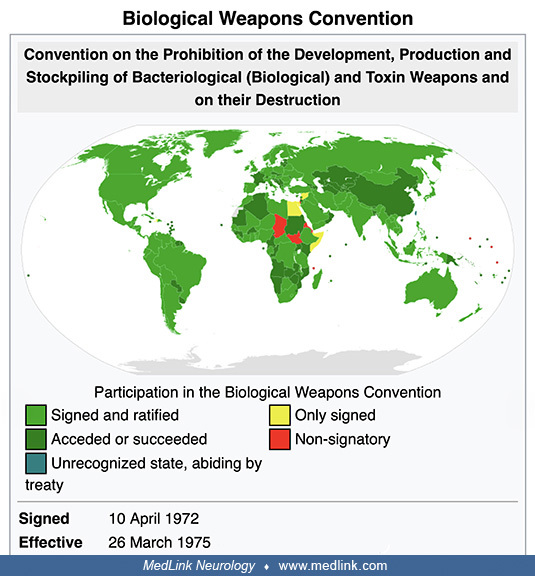
The Biological Weapons Convention (1972). Disarmament talks after World War II originally addressed biological and chemical weapons together but remained inconclusive. Shortly after states finalized negotiations on the Nuclear Non-Proliferation Treaty in 1968, an initiative in the United Kingdom broke the impasse in discussions of chemical and biological weapons: the UK proposed (1) to separate consideration of biological and chemical weapons and (2) to concentrate first on the former. From 1969 until 1971, the Biological Weapons Convention (BWC) was negotiated in Geneva, Switzerland, within the Eighteen Nation Committee on Disarmament (ENDC) and the Conference of the Committee on Disarmament (CCD).
Members of the Conference on Disarmament
Derivative work by User:NerdyNSK on September 13, 2008. Blank map by Users: Roke, Dbachmann, Hoshie, Wiz9999, Tene, Cp6, Nightstallion, NerdyNSK. (Courtesy of Wikimedia Commons. Creative Commons Attribution-Share Alike 3.0 Unpo...
Formally known as “The Convention on the Prohibition of the Development, Production, and Stockpiling of Bacteriological (Biological) and Toxin Weapons and on their Destruction,” the BWC opened for signature in 1972 and entered into force in March 1975.
The BWC prohibits developing, producing, acquiring, transferring, stockpiling, and using biological and toxin weapons. It was the first multilateral disarmament treaty banning an entire category of weapons of mass destruction. The BWC supplements the 1925 Geneva Protocol, which prohibited only the use of biological weapons.
|
Convention on the Prohibition of the Development, Production, and Stockpiling of Bacteriological (Biological) and Toxin Weapons and on Their Destruction ...
Article I. Each State Party to this Convention undertakes never in any circumstances to develop, produce, stockpile, or otherwise acquire or retain:
(1) microbial or other biological agents, or toxins whatever their origin or method of production, of types and in quantities that have no justification for prophylactic, protective, or other peaceful purposes;
(2) weapons, equipment, or means of delivery designed to use such agents or toxins for hostile purposes or in armed conflict.
(Convention On The Prohibition Of The Development, Production And Stockpiling Of Bacteriological (Biological) And Toxin Weapons And On Their Destruction. Accessed October 26, 2022.) |
Since 1980, States Parties have met approximately every 5 years to review the operation of the Biological Weapons Convention. The BWC has reached almost universal membership with 184 States Parties and four Signatory States (Convention on the Prohibition of the Development, Production and Stockpiling of Bacteriological (Biological) and Toxin Weapons and on Their Destruction. Accessed October 26, 2022). Unfortunately, the effectiveness of the BWC has been limited by insufficient institutional support and the absence of any formal verification process to monitor compliance.
-
A meeting of the Conference on Disarmament in the Council Chamber of the Palace of Nations
Photograph from U.S. Mission by Eric Bridiers on January 24, 2012. The United States Mission to the United Nations and Other International Organizations in Geneva represents the United States government in Geneva. (Creative Com...
-
Participation in the Biological Weapons Convention
Image by User:Allstar86 on July 29, 2008, most recently updated on August 14, 2019 (participation has been unchanged since then). (Creative Commons Attribution-Share Alike 3.0 Unported License, https://creativecommons.org/licen...
The 1984 Rajneeshee bioterrorism attack. In 1984, 751 people became ill with salmonellosis food poisoning in The Dalles, Oregon, due to deliberate Salmonella contamination of salad bars at 10 local restaurants. A group of prominent followers of Bhagwan Shree Rajneesh (later known as Osho, leader of the Rajneeshee cult), had hoped to incapacitate the voting population of the city so that their own candidates would win the 1984 Wasco County, Oregon, elections. The incident was the first and remains the single largest bioterrorist attack in United States history. The simplicity of the dissemination and its apparent effectiveness stimulated efforts by other terrorist groups with biological and chemical weapons.
-
Cult leader Osho Rajneesh
(Source: Wikimedia Commons. Image by Sjakkelien Vollebregt [1984]. Nationaal Archief, the Dutch National Archives. Creative Commons CC0 1.0 Universal Public Domain Dedication.)
-
Rajneesh and disciples at Poona, 1977
(Source: Wikimedia Commons. Image by Redheylin [1977]. Creative Commons CC0 1.0 Universal Public Domain Dedication.)
-
Rajneeshee cult members watch their leader, Osho Rajneesh
Members of the Rajneeshee cult watch their leader, Osho Rajneesh drive by in a car in the summer of 1982, 2 years before cult members perpetrated the largest bioterrorism event in United States history. (Source: © 2003 Samvado ...
-
Rajneeshee cult members, 1982 (1)
Rajneeshee cult members in the summer of 1982, 2 years before cult members perpetrated the largest bioterrorism event in United States history. (Source: © 2003 Samvado Gunnar Kossatz. Available at: web.archive.org/web/200710261...
-
Rajneeshee cult members, 1982 (2)
Rajneeshee cult members in the summer of 1982, 2 years before cult members perpetrated the largest bioterrorism event in United States history. (Source: © 2003 Samvado Gunnar Kossatz. Available at: web.archive.org/web/200710261...
-
Rajneeshee cult members, 1982 (3)
Rajneeshee cult members in the summer of 1982, 2 years before cult members perpetrated the largest bioterrorism event in United States history. (Source: © 2003 Samvado Gunnar Kossatz. Available at: web.archive.org/web/200710261...
-
Dutch sign suggesting that Rajneesh was a profiteer rather than a prophet, 1984
(Source: Wikimedia Commons. Image by Sjakkelien Vollebregt [1984]. Nationaal Archief, the Dutch National Archives. Creative Commons CC0 1.0 Universal Public Domain Dedication.)
-
Rajneeshee cult compound in The Dalles, Oregon
The compound of the Rajneeshee cult in The Dalles, Oregon, 1982. (Source: © 2003 Samvado Gunnar Kossatz. Available at: web.archive.org/web/20071026130939/http://m31.de/ranch/index.html. Used with permission.)
-
Restaurants affected by the Rajneeshee cult bioterrorism incident in 1984
Four of the restaurants in The Dalles, Oregon, affected by the Rajneeshee cult bioterrorism incident in 1984. (Source: Wikimedia Commons. User: Cacophony [2008]. Creative Commons Attribution 3.0 Unported [CC BY 3.0] license, cr...
-
Restaurant salsa bar intentionally contaminated as part of the Rajneeshee cult bioterrorism attack in 1983
The salsa bar of The Dalles' Taco Time that had been intentionally contaminated as part of the Rajneeshee cult bioterrorism attack in 1983 in The Dalles, Oregon. (Source: Wikimedia Commons. User: Cacophony [2008]. Creative Comm...
Prior use of anthrax as biological warfare agent. Anthrax has been used on a limited basis as both biological warfare and a bioterrorism agent.
The history of bioterrorism—anthrax
Dr. Joanne Cono of the Centers for Disease Control and Prevention discusses anthrax. (Source: U.S. Centers for Disease Control and Prevention, Atlanta, Georgia. Parts of this video were adapted from "Biological Warfare and Terrori...
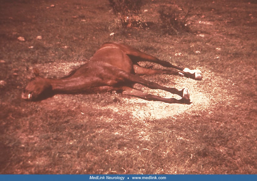
During World War I, German agents reportedly tried to infect allied horses as they were being shipped to the European front (41; 135), although other accounts discount this claim (114). Japan reportedly first developed anthrax as a bioweapon in the 1930s and used anthrax against China during World War II (114; 41; 61; 135; 126). In 1993, the Japanese terrorist group Aum Shinrikyo dispersed aerosols of anthrax and botulism throughout Tokyo on at least eight occasions (66; 67), but these attacks failed to produce illness, apparently in part because the terrorists mistakenly used a harmless anthrax strain developed for vaccines (106). In 2001, anthrax was used as a bioterrorism agent in the United States, with several deaths, more than 20 cases, and over 32,000 individuals receiving postexposure prophylaxis because of anthrax delivered through the United States mail system (23).
The United States began research on offensive bioweapons in 1943 at Camp Detrick (now Fort Detrick) in Frederick, Maryland. To assess the risk of covert biological attacks in the 1960s, the army conducted large-scale covert tests at various civilian sites (eg, National Airport and Greyhound Terminal in Washington, D.C., and the New York City subway), using the anthrax simulant Bacillus globigii (114). The United States bioweapons program, which included the development of weaponized anthrax, was terminated in 1969 following an executive order by President Richard Nixon. All stockpiles of biological agents were destroyed by May 1972 (126). There is no evidence that these U.S. weapons were ever deployed.
In 1972, the United States, the United Kingdom, and the USSR signed the Convention on the Prohibition of the Development, Production, and Stockpiling of Bacteriological (Biological) and Toxin Weapons and on Their Destruction ("The Biological Weapons Convention") (126). This treaty prohibits research, development, and stockpiling of biological agents for offensive military purposes. Although this biological weapons treaty has since been ratified by more than 140 countries, biological warfare research and the development of biological weapons continued in many countries (126).
The Soviet bioweapons program began before World War II under the auspices of Biopreparat, their bioweapons agency. This program continued until the 1990s despite the Biological Weapons Convention. By the 1980s, Biopreparat could produce thousands of tons of weaponized anthrax annually. Biopreparat also developed antibiotic-resistant anthrax using recombinant DNA techniques.
In April 1979, accidental release of anthrax spores occurred at a military bioweapons factory (Military Compound 19) in Sverdlovsk in the former Soviet Union (now Yekaterinburg, Russia) (01; 01b; 88; 69; 41; 132; 126). This incident resulted in at least 66 human deaths (among the 77 patients identified) in a narrow zone up to 4 kilometers downwind from the facility, as well as outbreaks of anthrax in livestock up to 50 kilometers downwind (88). Although the Soviet Ministry of Health initially blamed the deaths on cutaneous and gastrointestinal anthrax occurring from the consumption of contaminated meat, this scenario was doubted by military sources in the West (48; 94; 01; 88; 69; 41; 126). In 1992, Russian President Boris Yeltsin confirmed that this outbreak was a result of "military developments" (88), and clinical and epidemiologic studies documented inhalational anthrax from a mixture of different Bacillus anthracis strains as the cause (01; 88; 69).
In the aftermath of the Gulf War, Iraq acknowledged to the United Nations Special Commission Team 7 that it had conducted biological weapons research with various agents, including Bacillus anthracis (126). Further details were uncovered in 1995. Iraq had extensive research facilities at multiple sites, including Salman Park on the Tigris River. Iraq conducted field trials with Bacillus subtilis (an anthrax simulant) and various biowarfare agents using various delivery systems, including rockets, aerial bombs, sprayers attached to helicopters, and possibly unmanned drones (126). To reduce the particle size to maximize the delivered dose of anthrax, Iraq used sequential filters in an arrangement reportedly like that used at Camp Detrick in the 1950s. Iraq produced 8500 liters of concentrated anthrax, of which 6500 liters were placed into R400 bombs, Al Hussein warheads, and other devices (126).
Anthrax has been developed as a biological warfare agent by at least seven countries. It remains a significant bioweapons threat (47) and is considered the most likely biological warfare agent, as it is stable in spore form and can be stored for prolonged periods, it is easy and cheap to produce, there is no natural immunity in industrialized nations, it can be dispersed in air, the inhalational form is highly lethal, and the agent is difficult to detect (127). Nevertheless, aerosolized anthrax has not yet been used on the battlefield, possibly in part because of "moral repugnance" of biological weapons, potential retaliation in kind, delayed manifestations following use, and uncertain effects depending on weather conditions (107). Indeed, the uncertainties of such weapons are demonstrated by the Sverdlovsk incident in the USSR in 1979, which involved the accidental release of an estimated 10 kilograms of weapons-grade anthrax spores: this accident produced a total of only 66 fatalities among a potentially exposed population of 1.2 million people (107).
1979 Sverdlovsk anthrax outbreak in the Soviet Union
Dr. Matthew Meselson, Professor of Molecular and Cellular Biology at Harvard University, discusses the 1979 Sverdlovsk outbreak of anthrax in the Soviet Union, later determined to be the result of an accidental leak of anthrax spo...
Offensive anthrax bioweapons were initially manufactured as slurries of highly concentrated bacteria. Although easy and safe to manufacture, such slurries had to be refrigerated for storage and were difficult to disseminate (41). Later, freeze-dried powder formulations of anthrax spores were developed as bioweapons that, although technically difficult and dangerous to manufacture, were thermally stable without refrigeration and were easily disseminated (41). Bioweapon anthrax may also be modified to be multidrug-resistant and may be stabilized with other substances (eg, silica) to decrease electrostatic charges imparted by the milling process and, thereby, keep the particles from clumping. The latter issue is critical because biowarfare agents are most effectively delivered as an aerosol of particles from 1 to 5 microns in size (41). Particulates in this size range behave similarly to a gas and are taken into bronchioles and alveoli during respiration, whereas larger particles are much more difficult to disperse, rapidly fall to the ground, or become trapped in the upper airway (41). Anthrax aerosols are invisible, odorless, colorless, and tasteless.
Prior use of anthrax in bioterrorism. In late 2001, an anthrax outbreak attributed to bioterrorism occurred in the United States. Anthrax was spread through the mail (25; 26). Twenty-two cases were identified, 11 with inhalational anthrax and 11 with cutaneous anthrax (26; 27; 28; 29; 71; 68; 59). Five of the inhalational cases died. Only one of these cases had documented anthrax meningoencephalitis (71).
The FBI alleges that the 2001 anthrax bioterrorism outbreak in the United States was conducted by U.S. Army biodefense scientist Bruce Ivins, who worked at the U.S. Army Medical Research Institute of Infectious Diseases (USAMRIID) in Fort Dettrick, Maryland (90; 18). In 2008, shortly before these allegations were made public, Ivins committed suicide. The FBI employed a complex strategy to identify the source of the anthrax: (1) spores from the mailed envelopes were cultured, yielding thousands of colonies, one from each spore; (2) a small number of colonies with unusual features (“minority phenotypes”) were identified, and the genomes of these colonies were completely sequenced to identify the corresponding mutations; (3) tests were developed to screen anthrax samples for four of these mutations, using a polymerase chain reaction-based strategy; these molecular tests were applied to more than 1000 isolates from labs in the United States and other countries (56). In only eight of the study’s samples were all four mutations identified, and all were directly related to spores Ivins had created in 1997 (56). However, in February 2011, a scientific panel of the U.S. National Academy of Sciences independently evaluated the genetic evidence and concluded that it was insufficient to prove that Ivins was responsible. In addition, Ivins's laboratory did not contain the equipment needed to manufacture the refined powder of spores that were implicated in the 2001 bioterrorism attacks (18).
Anthrax is now classified as a category A biological warfare agent, a category of microorganisms or toxins that can be easily spread, leading to intoxication with potentially high death rates (06).
Prior attempts to use botulinum toxin in biowarfare and bioterrorism. Botulinum toxin has been considered by multiple countries as a potential bioweapons agent, with a possible attempt to use botulinum toxin by supporters of Francisco ("Pancho") Villa (1878-1923) against Mexican federal troops in 1910.
The history of bioterrorism--botulism
Dr. Joanne Cono of the Centers for Disease Control and Prevention discusses botulism. (Source: U.S. Centers for Disease Control and Prevention, Atlanta, Georgia. Parts of this video were adapted from "Biological Warfare and Terror...
During World War II, the Nazis reportedly developed botulinum toxin as a bioweapons agent. The United States and the Soviets also explored botulinum toxin as a potential bioweapon, both ultimately dismissing botulinum toxin as inferior to other agents such as anthrax and tularemia.
Saddam Hussein (1937-2006) began an extensive biological weapons program in Iraq in the early 1980s despite having signed (though not ratified until 1991) the Biological Weapons Convention (1972). Details of the Iraq bioweapons program surfaced after the Gulf War (1990-91) during the disarmament of Iraq under the United Nations Special Commission (UNSCOM). By the end of the war, Iraqi scientists had investigated the bioweapons potential of five bacterial strains, one fungal strain, five types of virus, and four toxins; of these, anthrax, botulinum toxin, and aflatoxin had proceeded to weaponization for possible deployment. Iraq claimed it had produced 19,000 liters of the toxin, sufficient (if true) to kill the world's entire human population. A vial of Clostridium botulinum bacteria was found by investigators in Iraq after the ouster of Hussein, leading to much greater public attention on the potential of botulinum toxin as a bioweapon.
In the 1990s, the Japanese doomsday cult Aum Shinrikyo produced botulinum toxin and spread it as an aerosol in downtown Tokyo, but the attacks caused no fatalities (82; 04; 05; 119).
In April 1990, cult members sprayed what they mistakenly believed was botulinum toxin from three trucks as they drove near two U.S. naval bases, Narita International Airport (one of two international airports serving the Greater Tokyo Area; also known as Tokyo-Narita, and formerly and originally known as New Tokyo International Airport), the National Diet (the National Legislature of Japan), the Imperial Palace, and the headquarters of a rival religious group (04). The goal was partly retaliation for the defeat of cult members who were running for political office and partly an attempt to precipitate an apocalyptic war.
On June 9, 1993, cult members sprayed what they mistakenly believed was botulinum toxin from a car equipped with a spraying device as it drove in Tokyo. The goal was to disrupt Prince Naruhito's wedding, seize power, and place the blame on the United States.
In the fall of 1994, cult members attempted to kill an attorney, Taro Takimoto, by mixing what they mistakenly believed was botulinum toxin into his drink. Takimoto had been assisting cult members who wished to leave the group and had been working on behalf of Aum victims.
On March 15, 1995, cult members attempted to spread what they mistakenly believed was botulinum toxin in the Kasumigaseki, Tokyo, subway station by placing briefcases equipped with custom spraying devices near the ticket barriers. The presumptive toxin had been replaced with a nontoxic substance (possibly water) by a dissident cult member. In any case, as in the cult's prior efforts, the cult had failed to acquire the necessary strain of Clostridium botulinum from which the botulinum toxin is derived.
Clinical manifestations
Presentation and course
Involvement of the nervous system is uncommon with most microorganisms that might reasonably be employed in biological warfare and bioterrorism.
Bacillus anthracis. Severe hemorrhagic meningitis regularly occurs with anthrax infection, often accompanied by severe systemic disease, most likely transmitted by spore inhalation.
All forms of anthrax infection (ie, cutaneous, gastrointestinal, inhalational, and injection) can be complicated by meningitis. With natural anthrax, anthrax bacilli most commonly enter the body via the skin and disseminate to the CNS via the hematogenous or lymphatic routes, leading to fatal bacterial meningitis. The interval from exposure to presentation of cardinal signs (ie, fever, dyspnea, and headache) varies from 2 to 60 days.
Botulinum toxin. Neurologists are most likely to become involved in primarily diagnosing bioterrorist attacks that use botulinum toxin, either through oral ingestion or inhalation (53). The characteristic descending paralysis typically starts in the extraocular and bulbar muscles, with associated autonomic features. Repetitive nerve stimulation usually shows an incremental muscle response. The differential diagnosis is from naturally occurring paralyzing illnesses (eg, Guillain-Barré syndrome, myasthenic crisis, diphtheria, tetrodotoxin, saxitoxin, snake envenomation, and from chemical warfare poisoning by organophosphates). Treatment is supportive.
Bioterrorists are more likely to contaminate victims’ inhaled air rather than the food supply. No immediate effects are noted after inhalation of botulinum toxin as the latent period varies from 1 to 5 days. The clinical features are predominantly a combination of multiple cranial nerve palsies and skeletal muscular paralysis. The early manifestations of botulin intoxication are usually ophthalmological, with symptoms such as blurred vision, diplopia, and ptosis. Signs include nystagmus, mydriasis, and extraocular muscle dysfunction. Other symptoms, such as dysphagia and dysarthria, are associated with descending flaccid paralysis that typically develops 12 to 72 hours after exposure. The autonomic effects of botulism are manifested by anticholinergic signs and symptoms (eg, dry mouth, ileus, constipation, and urinary retention). Sensory symptoms are absent. Because botulinum toxins do not cross the blood-brain barrier and do not affect the brain, affected individuals are usually alert and oriented. With severe respiratory muscle paralysis, the patient may become cyanotic or exhibit narcosis from CO2 retention. Respiratory paralysis may lead to death.
Prognosis and complications
Anthrax. Untreated, the case fatality from cutaneous anthrax is about 20% to 30%, compared to 25% to 60% with gastrointestinal anthrax, and nearly 100% with inhalational anthrax. With treatment, mortality is less than 1% with cutaneous anthrax (41; 51; 135; 62), whereas mortality remains high, even with aggressive treatment, with gastrointestinal or inhalational anthrax (64; 62). Inhalational anthrax was generally thought to be fatal in 80% to over 90% of cases (100; 14; 01; 135), but the mortality in the 2001 U.S. outbreak has been much better than anticipated (5 of 11 cases or 45%) with the use of aggressive treatment and intensive care unit support, including mechanical ventilation and dialysis as necessary (23; 135). From 2001 to 2014, 8 of 15 (53%) patients with inhalation anthrax survived with early diagnosis, combination antimicrobial drug treatment to eradicate the bacteria and inhibit toxin production, and aggressive pleural effusion management (62). Initiation of antibiotic or anthrax antiserum therapy during the prodromal phase of inhalational anthrax is associated with markedly improved survival compared with initiation of treatment of fulminant cases (64). Nevertheless, anthrax survivors report significant health problems and poor life adjustment 1 year after the onset of bioterrorism-related anthrax infection (108).
Anthrax meningoencephalitis is usually rapidly fatal, with roughly two-thirds of affected patients dying within 24 hours of presentation (80; 95), although there are a small number of reported survivals following anthrax meningoencephalitis (45; 111; 129; 121; 123; 75; 120; 80; 09). In a systematic review, survival of anthrax meningitis was predicted by treatment with a bactericidal agent and the use of multiple antimicrobials (72). There is limited information on long-term outcomes among the few survivors, but several cases were reported to have fully recovered (121; 120; 80; 09).
Botulism. The advent of modern supportive care has improved botulism prognosis so that the case fatality rate for food-borne botulism has decreased from the 50% to 70% range to the 5% to 20% range. Although botulinum neurotoxins bind irreversibly to presynaptic endplates and impair them irreparably, axons can regenerate new endplates, and full recovery can occur, although it may take several months. The case fatality and outcomes from biological warfare or terrorism could easily be much worse than the results obtained with individual sporadic cases or cases from small natural outbreaks because the medical capacity could be overwhelmed and treatment supplies (eg, Botulism Antitoxin Heptavalent [A, B, C, D, E, F, G]) could be exhausted by sudden widescale demand.
Biological basis
Etiology and pathogenesis
Various agents used for biological and chemical warfare are listed in Table 1. Many of them can cause neurologic manifestations. Encephalomyelitic viruses, toxins, and chemicals are more likely to be associated with neurotoxicity. Some of the bacterial infections may be associated with neurologic complications.
Characteristics that make a pathogen a high risk for bioterrorism include a low infective dose, ability to be aerosolized, high contagiousness, and hardiness (ie, survival in various environmental conditions) (03).
Table 1. Potential Biological Warfare or Bioterrorism Agents with Frequent Involvement of the Nervous System
Bacteria |
• Bacillus anthracis (inhalation anthrax) • Clostridium botulinum |
Viruses |
• Eastern equine encephalitis • Venezuelan equine encephalitis • Western equine encephalitis |
Toxins |
• Botulinum • Brevetoxins • Conotoxins • Saxitoxins • Tetrodotoxin |
CDC categorization of bioterrorism agents and diseases. The Centers for Disease Control and Prevention (CDC) has a 3-tier categorization system for bioterrorism agents and diseases (CDC: Bioterrorism Agents and Diseases):
Category A. Category A is comprised of high-priority agents that pose a risk to national security because (1) they can be easily disseminated or transmitted from person to person, (2) they have the potential for major public health impact (eg, high case-fatality rates), (3) they will likely cause public panic and social disruption, and (4) they require special measures for public health preparedness. The two Category A diseases with the greatest potential to affect the nervous system as a major feature of the illness are anthrax (Bacillus anthracis) and botulism (Clostridium botulinum toxin). Other Category A diseases or agents include plague (Yersinia pestis), smallpox (variola major), tularemia (Francisella tularensis), and viral hemorrhagic fevers (eg, Ebola, Marburg) (06; 07).
Category B. Category B is comprised of moderate-priority agents that (1) are moderately easy to disseminate, (2) result in moderate morbidity rates and low mortality rates, and (3) require specific enhancements for diagnostic capacity and warrant enhanced disease surveillance. Of these, only viral encephalitis viruses (alphaviruses, such as eastern equine encephalitis, Venezuelan equine encephalitis, and western equine encephalitis) are likely to primarily affect the nervous system (112).
-
Hypoattenuation of left caudate head and diencephalic region in eastern equine encephalitis (CT)
Noncontrast CT scan of the brain of a 5-year-old girl with eastern equine encephalitis on hospital day 2 showing subtle hypoattenuation of the left caudate head (arrow) and diencephalic region. (Source: Silverman MA, Misasi J, ...
-
Hyperintensities in bimesial temporal and dorsal pontomesencephalic regions in eastern equine encephalitis (MRI)
Axial FLAIR image from a brain MRI scan of a 13-year-old boy with eastern equine encephalitis on hospital day 2 showing abnormal T2 hyperintense regions of the bimesial temporal regions (thick arrows) with accompanying abnormal...
-
Hyperintenities in caudate and thalamic nuclei in eastern equine encephalitis (MRI)
Axial FLAIR from a brain MRI scan of a 3.5-year-old girl with eastern equine encephalitis on hospital day 3 showing abnormal T2 hyperintense caudate and thalamic nuclei, most prominent on the right (arrow). (Source: Silverman M...
-
Hyperintensities in parietotemporal gray matter and subcortical white matter in eastern equine encephalitis (MRI)
Axial FLAIR from a brain MRI scan of a 3.5-year-old girl with eastern equine encephalitis on hospital day 3. The abnormal T2 hyperintense regions are most prominent in the right parietotemporal gray matter (arrow) and subcortic...
-
Histopathologic features in eastern equine encephalitis (temporal lobe)
Histopathologic features from a 5-year-old girl with eastern equine encephalitis in 2005, as part of a study of eastern equine encephalitis in Massachusetts and New Hampshire from 1970 to 2010. The postmortem samples of central...
-
Histopathologic features in eastern equine encephalitis (midbrain)
Histopathologic features from a 5-year-old girl with eastern equine encephalitis in 2005, as part of a study of eastern equine encephalitis in Massachusetts and New Hampshire from 1970 to 2010. The postmortem samples of central...
-
Histopathologic features in eastern equine encephalitis (basal ganglia) (1)
Histopathologic features from a 5-year-old girl with eastern equine encephalitis in 2005, as part of a study of eastern equine encephalitis in Massachusetts and New Hampshire from 1970 to 2010. The postmortem samples of central...
-
Histopathologic features in eastern equine encephalitis (basal ganglia) (2)
Histopathologic features from a 5-year-old girl with eastern equine encephalitis in 2005, as part of a study of eastern equine encephalitis in Massachusetts and New Hampshire from 1970 to 2010. The postmortem samples of central...
-
Histopathologic features in eastern equine encephalitis (thalamus)
Histopathologic features from a 5-year-old girl with eastern equine encephalitis in 2005, as part of a study of eastern equine encephalitis in Massachusetts and New Hampshire from 1970 to 2010. The postmortem samples of central...
-
Immunohistochemistry demonstrating eastern equine encephalitis viral antigens in the thalamus
Histopathologic features from a 5-year-old girl with eastern equine encephalitis in 2005, as part of a study of eastern equine encephalitis in Massachusetts and New Hampshire from 1970 to 2010. The postmortem samples of central...
-
Eastern equine encephalitis virus neuroinvasive disease cases reported by year, 2011-2020
(Source: ArbotNet, Centers for Disease Control and Prevention, Atlanta, Georgia. Public domain.)
-
Eastern equine encephalitis virus neuroinvasive disease average annual incidence by county of residence, 2011-2020
(Source: ArbotNet, Centers for Disease Control and Prevention, Atlanta, Georgia. Public domain.)
-
Eastern equine encephalitis virus neuroinvasive disease cases reported by state of residence, 2011-2020
(Source: ArbotNet, Centers for Disease Control and Prevention, Atlanta, Georgia. Public domain.)
Category C. Category C is comprised of lower-priority agents, including emerging pathogens that could potentially be engineered for mass dissemination because of availability, ease of production and dissemination, or potential for major health impact (eg, potential for high morbidity or mortality).
Bacillus anthracis (Category A). Insight regarding the mechanism for the spread of B anthracis by inhalation is provided by pathologic studies of victims from the Sverdlovsk epidemic of 1979. The contagion mechanism was traced to the release of aerosols containing this organism at a secret biological-agent production facility in Russia (60).
Accidental release of anthrax from an illegal biological warfare facility in Sverdlovsk, Russia, in 1979
(Source: U.S. National Archives [1984]. Combined Military Service Digital Photographic Files. Released to the public. Public domain.)
A hematogenous spread involved the central nervous system with resulting vasculitis, hemorrhages, and edema due to the toxins. The risk of hemorrhagic meningitis after exposure to anthrax inhalation is estimated to be as high as 50% (66; 67).
Histopathology in case of fatal human anthrax with hemorrhagic meningitis
Note the rod-shaped, darkly stained, Bacillus anthracis bacteria. Photomicrograph by CDC/Dr. Marshall Fox in 1976. (Source: Public Health Image Library, U.S. Centers for Disease Control and Prevention, Atlanta, Georgia...
Equine encephalitis (Category B). Venezuelan equine encephalitis virus could be developed for transmission by inhalation with secondary zoonotic transmission cycles sustained by horses and mosquitoes (53), as could eastern and western equine encephalitis viruses.
The Eastern equine encephalitis virus is spread to people by the bite of an infected mosquito (15).
-
Eastern equine encephalitis virus particles in mosquito salivary gland tissue (1)
Transmission electron microscopic image reveals numerous Eastern equine encephalitis virus particles harbored in this mosquito salivary gland tissue specimen. Photomicrograph by CDC/Dr. Fred Murphy and Sylvia Whitfield in 1975....
-
Eastern equine encephalitis virus virions in central nervous system tissue (1)
Transmission electron microscopic image reveals numerous Eastern equine encephalitis virus virions in this central nervous system tissue specimen. Photomicrograph by CDC/Dr. Fred Murphy and Sylvia Whitfield in 1975. (Source: Pu...
-
Eastern equine encephalitis virus virions in tissue specimen (1)
Negatively stained transmission electron microscopic image reveals Eastern equine encephalitis virus virions in this tissue specimen. Photomicrograph by CDC/Dr. Fred Murphy and Sylvia Whitfield in 1975. (Source: Public Health I...
-
Eastern equine encephalitis virus virions in central nervous system tissue (2)
Transmission electron microscopic image reveals numerous Eastern equine encephalitis virus virions in this specimen of central nervous system tissue. Photomicrograph by CDC/Dr. Fred Murphy and Sylvia Whitfield in 1975. (Source:...
-
Eastern equine encephalitis virus virions in central nervous system tissue (3)
Transmission electron microscopic image reveals numerous Eastern equine encephalitis virus virions in this specimen of central nervous system tissue. Photomicrograph by CDC/Dr. Fred Murphy and Sylvia Whitfield in 1975. (Source:...
-
Eastern equine encephalitis virus virions in tissue specimen (2)
Transmission electron microscopic image reveals Eastern equine encephalitis virus virions in this tissue specimen. Photomicrograph by CDC/Dr. Fred Murphy and Sylvia Whitfield in 1975. (Source: Public Health Image Library, U.S. ...
-
Salivary gland tissue section extracted from a mosquito infected by the eastern equine encephalitis virus
In this digitally colorized transmission electron microscopic image, the viral particles have been colorized red (magnification x 83,900) Photomicrograph by CDC/Dr. Fred Murphy and Sylvia Whitfield in 1975. (Source: Public Heal...
-
Ultrastructural features exhibited by gut cells of an Aedes triseriatus mosquito infected with Eastern equine encephalitis virus
In this transmission electron microscopic image, note the numerous Eastern equine encephalitis virions in various stages of development, from the precursor particles through the mature virus. Image by CDC, 1969. (Source: Public...
In the United States, most cases occur in eastern or Gulf Coast states.
Reported human cases of Eastern equine encephalitis from 1964-2010
(Source: ArboNET, Arboviral Diseases Branch, Centers for Disease Control and Prevention. Public domain.)
Although rare, approximately 30% of people with Eastern equine encephalitis die, and many survivors have residual neurologic problems. Eastern equine encephalitis, commonly called Triple E or sleeping sickness (not to be confused with African trypanosomiasis), is caused by a zoonotic mosquito-vectored Togavirus that is present in North, Central, and South America and the Caribbean.
-
Culiseta melanura mosquito (dorsal view)
The diagram illustrates characteristics that include a long, curved proboscis, a dark-scaled abdomen, and slightly enlarged, dark scales on the outer wing. This mosquito is a vector of the eastern equine encephalitis virus in b...
-
Uranotaenia sapphirina mosquito (right lateral view)
This image depicts a Uranotaenia sapphirina mosquito, along with an accompanying inset of its right wing from a superior perspective, detailing its venation pattern. Known as the sapphire-lined mosquito, U. sapphirina gets its ...
-
Culiseta melanura mosquito preparing to feed on a blue jay
Eastern equine encephalitis virus is maintained in a cycle between C. melanura mosquitoes and avian hosts in freshwater hardwood swamps. C. melanura is not an important vector of Eastern equine encephalitis vi...
In the United States, Eastern equine encephalitis is often associated with coastal plains, most commonly in East Coast and Gulf Coast states. No human vaccines or licensed therapeutic drugs are available in the U.S. for Eastern equine encephalitis. Eastern equine encephalitis virus is a potential bioweapon, particularly because it is transmissible by aerosol.
The Western equine encephalomyelitis virus is an alphavirus of the family Togaviridae.
Cryogenic electron microscopy model of Western equine encephalitis virus at 12Å resolution
(Source: User: A2-33 on December 3, 2013. Courtesy of Wikimedia Commons. Creative Commons Attribution-Share Alike 3.0 Unported License, https://creativecommons.org/licenses/by-sa/3.0/deed.en.)
The Western equine encephalomyelitis virus is an arbovirus (arthropod-borne virus) transmitted by mosquitoes of the genera Culex and Culiseta.
-
Culex tarsalis mosquito (close anterior view)
In this photograph, the Culex tarsalis mosquito is about to begin feeding after landing on the skin of what would become its human host. Note the light-colored band wrapped around its dark-scaled proboscis (A), the mul...
-
Culex tarsalis mosquito (dorsal view)
Culex tarsalis mosquito is a vector of many mosquito-borne diseases. The epidemiologic importance of C. tarsalis lies in its ability to spread Western equine encephalitis, St. Louis encephalitis, and Californi...
The virus is transmitted to people and horses by bites from infected mosquitoes (Culex tarsalis and Aedes taeniorhynchus), particularly during wet summer months; birds are the natural reservoir of the virus.
Western equine encephalitis virus cycle in Western United States
This diagram illustrates the methods by which the arbovirus Western equine encephalitis virus reproduces and amplifies itself in both avian and rodent populations and is subsequently transmitted by the Culex tarsalis m...
In North America, Western equine encephalomyelitis occurs primarily in U.S. states and Canadian provinces west of the Mississippi River. The disease also occurs in South America. Western equine encephalomyelitis is commonly a subclinical infection in adult humans, but the disease can cause serious sequelae in infants, children, and the elderly. The overall mortality of Western equine encephalomyelitis is low (approximately 4%) and is associated mostly with infection in the elderly. Approximately 15% to 20% of horses that acquire the virus die or need to be euthanized.
Horse infected by mosquito-borne Western equine encephalitis virus
Note the characteristic head droop. Photograph by CDC/James Stewart, Fort Collins in 1976. (Source: Public Health Image Library, U.S. Centers for Disease Control and Prevention, Atlanta, Georgia. Image 2625. Public domain.)
No human vaccines or licensed therapeutic drugs are available in the U.S. for Western equine encephalomyelitis. Western equine encephalitis virus was one of more than a dozen agents the United States researched as potential biological weapons before the nation suspended its biological weapons program.
Venezuelan equine encephalitis is a mosquito-borne disease endemic in regions of Central and South America that causes sporadic outbreaks of equine and human encephalitis. The virus induces neural necrosis and edema in affected mammals.
-
Dead horse that was a victim of a Venezuelan equine encephalitis outbreak in Texas
As in humans, under natural conditions (as opposed to the artificial condition of bioweapons use), the etiologic pathogens responsible for equine encephalitic diseases are transmitted to horses by mosquito vectors, with various...
-
Histopathologic changes indicative of neural necrosis and edema in Venezuelan equine encephalitis (1)
This photomicrograph of a mouse brain tissue sample collected after the mouse had succumbed to Venezuelan equine encephalitis reveals histopathologic changes indicative of neural necrosis and edema. Photomicrograph by CDC/Dr. F...
-
Histopathologic changes indicative of neural necrosis and edema in Venezuelan equine encephalitis (2)
This photomicrograph of a mouse brain tissue sample collected after the mouse had succumbed to Venezuelan equine encephalitis reveals histopathologic changes indicative of neural necrosis and edema (magnification x 300). Photom...
-
Histopathologic changes indicative of neural necrosis and edema in Venezuelan equine encephalitis (3)
This photomicrograph of a mouse brain tissue sample collected after the mouse had succumbed to Venezuelan equine encephalitis reveals histopathologic changes indicative of neural necrosis and edema (magnification x 300). Photom...
-
Histopathologic changes indicative of neural necrosis and edema in Venezuelan equine encephalitis (4)
This photomicrograph of a mouse brain tissue sample collected after the mouse had succumbed to Venezuelan equine encephalitis reveals histopathologic changes indicative of neural necrosis and edema (magnification x 300.) Photom...
-
Histopathologic changes indicative of neural necrosis and edema in Venezuelan equine encephalitis (5)
This photomicrograph of a mouse brain tissue sample collected after the mouse had succumbed to Venezuelan equine encephalitis reveals histopathologic changes indicative of neural necrosis and edema (magnification x 300). Photom...
-
Venezuelan equine encephalitis virus virions in tissue specimen fixed using phosphotungstic acid
This negatively stained, transmission electron microscopic image revealed Venezuelan equine encephalitis virus virions in the tissue specimen, which had been fixed using phosphotungstic acid. Phosphotungstic acid is very electr...
Toxins. The term “toxin” refers to a toxic substance of biological origin, although simple toxins can be synthesized in the laboratory or produced by genetic modification in other species.
Bacterial toxins are of two general types: proteinaceous exotoxins (eg, tetanus, diphtheria, or botulinum toxins) are part of a defensive system of bacteria (to avoid capture, ingestion, etc.) and so are typically secreted into the surrounding medium, in contrast to endotoxins (eg, lipopolysaccharides in the outer membrane of gram-negative bacteria) that are only released when the bacterium disintegrates.
Mycotoxins are diverse and could be used by terrorist organizations to poison food and water sources, although dispersal in indoor air appears to be the most effective method for a bioterrorist attack (84). Crude concentrated or dried extracts of readily grown fungal cultures can simultaneously produce several toxins with synergistic actions, but mycotoxin bioterror weapons are more likely to involve the liver (eg, aflatoxins, fumonisin B1), kidneys (ochratoxin A, fumonisin B1), lungs (fumonisin B1), gastrointestinal tract (trichothecenes type A, eg, T-2 toxin; type B, eg, deoxynivalenol [DON]), and bone marrow (trichothecenes type A, eg, T-2 toxin) than the nervous system (83; 63; 115; 96; 84; 70).
As chemical products of living organisms, toxins, when employed by people for a nefarious purpose, occupy an ill-defined “no man’s land” between chemical warfare and biological warfare agents. From a pharmacological and toxicological point of view, toxins could be considered chemical weapons, but most experts and the U.S. Army classify toxins as biological weapons.
Toxins most relevant to bioterrorism include ricin, botulinum, Clostridium perfringens epsilon toxin, conotoxins, Shiga toxins, saxitoxins, tetrodotoxins, mycotoxins, nicotine (03), and brevetoxin. Of these, botulinum, brevetoxin, conotoxins, nicotine, saxitoxins, and tetrodotoxin are most likely to involve the nervous system.
(1) Botulinum toxin (Category A). It is feasible to deliver botulinum toxin as an aerosolized biological weapon. Botulinum toxins are a series of seven related protein neurotoxins, serotypes A through G.
Botulinum neurotoxin serotype A produced by Clostridium botulinum (3D ribbon model)
(Source: Wikimedia Commons. Created on October 4, 2008. See also: Lacy DB, Tepp W, Cohen AC, DasGupta BR, Stevens RC. Crystal structure of botulinum neurotoxin type A and implications for toxicity. Nat Struct Biol 1998;5[10]:89...
Each serotype is a protein of approximately 150 kD produced by a separate strain of the gram-positive anaerobic bacterium Clostridium botulinum. Botulinum toxins block acetylcholine release from the presynaptic nerve terminal in the peripheral nervous system, leading to muscle paralysis. Botulinum toxin A is the most toxic serotype for humans. It can be readily aerosolized and persists in this state for weeks in still water and food but degrades on exposure to heat, acidity, or air for more than 12 hours.
Botulinum toxin binds to high-affinity recognition sites on the cholinergic nerve terminals (there may be a separate one for each serotype). The toxin enters the nerve terminal by receptor-mediated endocytosis but then is extruded into the cytoplasm. In the cytoplasm, the toxin cleaves SNARE proteins (ie, proteins that mediate vesicle fusion with the presynaptic membrane), preventing the cell from releasing vesicles of neurotransmitter, interrupting neurotransmission, and causing paralysis (55; 38).
-
Mechanism of action of botulinum toxins
Synaptobrevin on the synaptic vesicle must interact with syntaxin and SNAP (synaptosomal-associated protein)-25 on the neuronal membrane for fusion to occur, which allows the nerve impulse to be delivered across the synaptic ju...
-
Molecular targets of clostridial neurotoxins in the presynaptic axon terminal of a neuron
Abbreviations: BoNT/A-G=botulinum toxin A-G, TeNT=tetanus toxin. (Source: Y tambe on June 26, 2007. Commons Attribution-Share Alike 3.0 Unported License, https://creativecommons.org/licenses/by-sa/3.0/deed.en.)
Botulinum toxin consists of two polypeptide subunits: A and B chains. The B subunit binds to receptors on the axons of motor neurons. The toxin is taken into the axon by receptor-mediated endocytosis, and then the A chain acts to prevent fusion of the synaptic vesicle with the cell membrane, thus, preventing the release of acetylcholine. This mechanism of blocking neuromuscular transmission is called presynaptic inhibition. The presynaptic inhibition interrupts both cholinergic autonomic (muscarinic) and motor (nicotinic) neurotransmission, producing the cranial neuropathies and skeletal muscle paralysis seen in clinical botulism. Recovery only occurs after the neuron generates a new axon terminal, which can take months.
The estimated lethal dose of type A toxin is 1.3 to 2.1 ng/kg intravenously or intramuscularly, 10 to 13 ng/kg when inhaled, or 1000 ng/kg orally.
(2) Brevetoxins (Category C). Brevetoxins are a group of tasteless and odorless neurotoxic cyclic polyether compounds produced naturally by species of dinoflagellates (Karenia sp.). Brevetoxins bind to voltage-gated sodium channels in nerve cells, leading to disruption of normal neurologic processes and causing neurotoxic shellfish poisoning. The typical natural route of human exposure is by ingestion of contaminated shellfish. Ingestion of these toxins produces a combination of gastrointestinal and neurologic signs and symptoms; gastrointestinal symptoms include abdominal pain, vomiting, and diarrhea, and the most disabling neurologic signs and symptoms include vertigo and ataxia. Inhalational exposure may cause respiratory symptoms, such as cough, dyspnea, and bronchospasm. Toxicity can also result from dermal exposure.
(3) Conotoxins (Category C). Conotoxins, the venom of various species of cone snails, are pharmacologically and chemically diverse.
-
3D structure of alpha-conotoxin PnIB from Conus pennaceus
The schematic representation is colored from N-terminal end (blue) to C-terminal end (red). Disulfide bonds are shown in yellow. (Source: Created by Fvasconcellos on June 26, 2008. Released into Public domain.)
-
Molecular structure of Mu-conotoxin pIIIa
Cartoon representation. (Source: Jawahar Swaminathan and MSD staff at the European Bioinformatics Institute. Released into public domain. Figure edited by Dr. Douglas J Lanska.)
-
3D structure of omega-conotoxin MVIIA (ziconotide)
The schematic representation is colored from N-terminal end (red) to C-terminal end (blue). Disulfide bonds are shown in gold. (Source: Created by Fvasconcellos on June 29, 2008. Released into Public domain.)
Conotoxins affect neurotransmitter receptors (eg, nicotinic, adrenergic, NMDA, and serotonergic) and ion channels (eg, sodium, potassium, and calcium).
-
Eukaryotic voltage-gated sodium channels and the outer pore blockers: 3D structures of the outer pore-blocking toxins
Shown is Arg13 in μ-conotoxin GIIIA, which is critical for the channel block. (Source: Tikhonov DB, Zhorov BS. Predicting structural details of the sodium channel pore basing on animal toxin studies. Front Pharmacol 2018;9:880....
-
Coupled movement of a sodium ion and ligand QX-222 in the closed cardiac sodium channel (3)
A μ-conotoxin (μ-CTX) mutant does not completely block the sodium current. The sodium ion can escape through the incompletely sealed outer pore and vacate the cation-attractive central cavity for binding of QX-222, which is a l...
The most lethal effect of conotoxins to humans is diaphragmatic paralysis and respiratory arrest. Most conotoxins are not a bioterrorism threat, but some could be weaponized and used as an aerosol (eg, a-conotoxins, k-conotoxins, and o-conotoxins).
(4) Saxitoxins (Category C). More than 50 structurally related neurotoxins (known collectively as "saxitoxins") are produced by protists, algae, and cyanobacteria. Saxitoxin, the best-known paralytic shellfish toxin, is one of the most potent known natural toxins. Saxitoxin acts as a selective, reversible, voltage-gated sodium channel blocker. Saxitoxin binds directly in the pore of the channel protein, occluding the opening, and preventing the flow of sodium ions through the membrane.
(5) Tetrodotoxin (Category C). Tetrodotoxin is an extremely potent toxin naturally found mainly in the liver and sex organs (gonads) of some fish (eg, puffer fish, globefish, and toadfish) and in some amphibian, octopus, and shellfish species.
Structural chemical formula for tetrodotoxin
Tetrodotoxin binds to the voltage-gated sodium channels in nerve cell membranes and blocks the passage of sodium ions (responsible for the rising phase of an action potential) into the neuron (81). This mechanism of action was established in 1964 by Toshio Narahashi (1927-2013), John W Moore (1920-2019), and William R Scott at Duke University (92; 91).
-
Blockade of tetrodotoxin-sensitive voltage-gated sodium channels
The normal condition is depicted in the top image, and the tetrodotoxin blockade (black blobs on the outer surface of the voltage-gated sodium channels) is depicted in the lower image. TTX=tetrodotoxin. (Source: Campos-Ríos A, ...
-
Model of voltage-gated sodium channels: extracellular view of the tetrodotoxin-bound model
Repeats I, II, III, and IV are green, yellow, cyan, and magenta, respectively. The outer carboxylates are shown as sticks. (Source: Tikhonov DB, Zhorov BS. Predicting structural details of the sodium channel pore basing on anim...
-
Coupled movement of a sodium ion and ligand QX-222 in the closed cardiac sodium channel (1)
In the schematic, the permanently charged QX-222 reaches the inner pore via III/IV repeat interface and displaces the resident ion that leaves the central cavity through the selectivity filter. QX-222, a lidocaine derivative, i...
-
Coupled movement of a sodium ion and ligand QX-222 in the closed cardiac sodium channel (2)
When the outer pore is blocked by tetrodotoxin (TTX), the sodium ion is trapped in the central cavity and prevents binding of QX-222. QX-222, a lidocaine derivative, is a sodium channel blocker. (Source: Tikhonov DB, Zhorov BS....
Brevetoxins, saxitoxins, tetrodotoxin, and some conotoxins all act to block voltage-gated sodium channels.
-
Neurotoxin sodium channel binding sites
The diagram illustrates the membrane topology of a voltage-gated sodium channel protein. Binding sites for different neurotoxins are indicated by color. Abbreviations: TTX, tetrodotoxin; STX, saxitoxin. Image by Kathleen Cusick...
-
Eukaryotic voltage-gated sodium channels and the outer pore blockers: alpha-subunit transmembrane topology
Positions of the selectivity filter "DEKA residues" and the outer carboxylates, which interact with TTX (tetrodotoxin), STX (saxitoxin), and μ-CTX (μ-conotoxin), are marked by red dots. (Source: Tikhonov DB, Zhorov BS. Predicti...
-
Eukaryotic voltage-gated sodium channels and the outer pore blockers: general 3D model of sodium channels
Positions of toxin-interacting residues are shown by red circles. In eukaryotic voltage-gated sodium channels, the selectivity filter DEKA ring, which contains D, E, K, and A residues, borders the extracellularly exposed outer ...