Neuroimmunology
Hashimoto encephalopathy
May. 19, 2025
MedLink, LLC
3525 Del Mar Heights Rd, Ste 304
San Diego, CA 92130-2122
Toll Free (U.S. + Canada): 800-452-2400
US Number: +1-619-640-4660
Support: service@medlink.com
Editor: editor@medlink.com
ISSN: 2831-9125
Toll Free (U.S. + Canada): 800-452-2400
US Number: +1-619-640-4660
Support: service@medlink.com
Editor: editor@medlink.com
ISSN: 2831-9125
Nearly 3,000 illustrations, including video clips of neurologic disorders.
Every article is reviewed by our esteemed Editorial Board for accuracy and currency.
Full spectrum of neurology in 1,200 comprehensive articles.
Listen to MedLink on the go with Audio versions of each article.
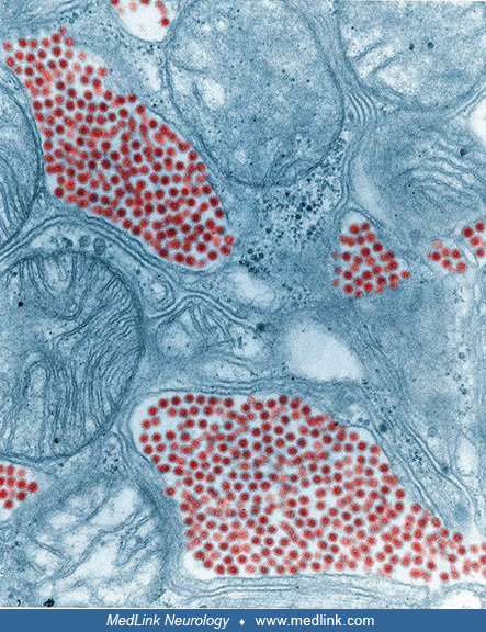
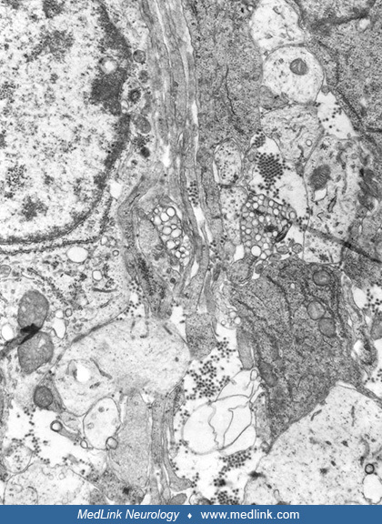
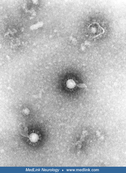
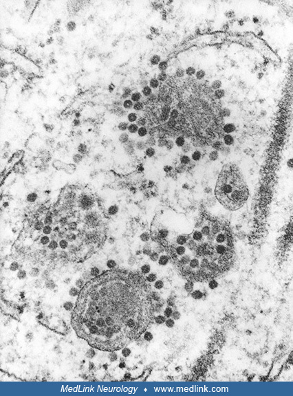
In this transmission electron microscopic image, note the numerous Eastern equine encephalitis virions in various stages of development, from the precursor particles through the mature virus. Image by CDC, 1969. (Source: Public Health Image Library, U.S. Centers for Disease Control and Prevention, Atlanta, Georgia. Image 17438. Public domain.)