Neuropharmacology & Neurotherapeutics
Suzetrigine
May. 14, 2026
MedLink, LLC
3525 Del Mar Heights Rd, Ste 304
San Diego, CA 92130-2122
Toll Free (U.S. + Canada): 800-452-2400
US Number: +1-619-640-4660
Support: service@medlink.com
Editor: editor@medlink.com
ISSN: 2831-9125
Toll Free (U.S. + Canada): 800-452-2400
US Number: +1-619-640-4660
Support: service@medlink.com
Editor: editor@medlink.com
ISSN: 2831-9125
Nearly 3,000 illustrations, including video clips of neurologic disorders.
Every article is reviewed by our esteemed Editorial Board for accuracy and currency.
Full spectrum of neurology in 1,200 comprehensive articles.
Listen to MedLink on the go with Audio versions of each article.
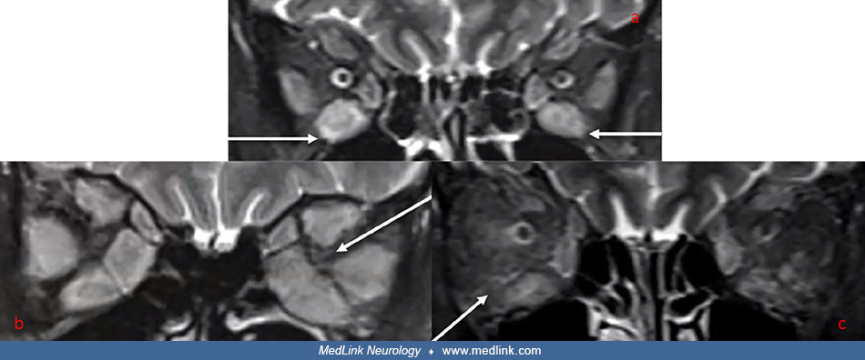
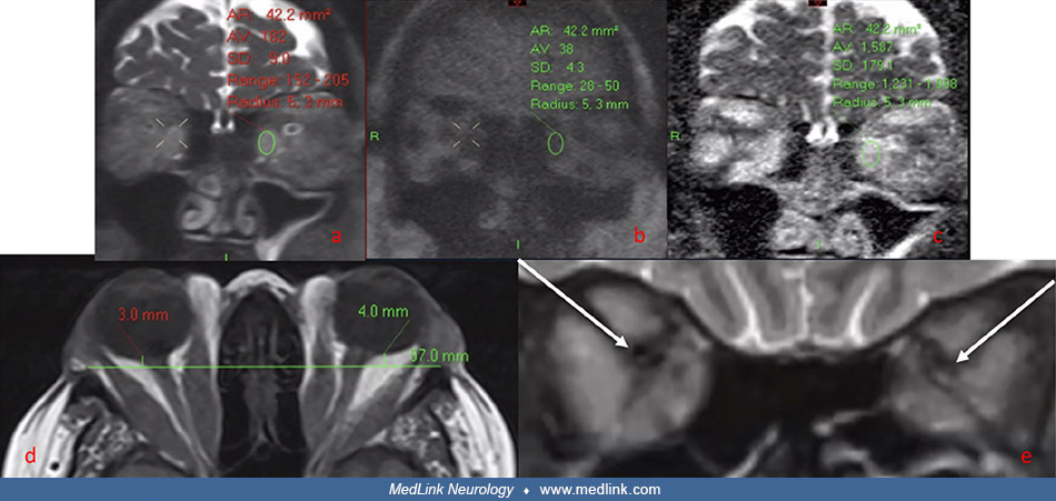
Coronal non‐EPI HASTE DWI MRI images ([a] = b0, [b] = b1000, [c] = ADC map) in a patient with severe thyroid eye disease. A region of interest has been placed in the left medial rectus muscle belly, showing markedly elevated ADC (1587) on (c). (d) Axial MRI T1 image, demonstrating radiological measurement of proptosis--the distance between the inter‐zygomatic line and the posterior globe in a patient with thyroid eye disease (normal range: 5.00 to 10.00 mm posterior to the inter‐zygomatic line). (e) Coronal MRI STIR image showing apical crowding (arrow) of the optic nerves bilaterally in a patient with thyroid eye disease and dysthyroid optic neuropathy. (From: Moledina M, Lee V, Bhatia K, et al. Radiological Activity Score [RAS]-MRI characteristics in dysthyroid optic neuropathy in a multi-ethnic thyroid eye disease population. Clin Endocrinol [Oxf] 2025;103[3]:385-95. Creative Commons Attribution 4.0 International [CC BY 4.0] license, creativecommons.org/licenses/by/4.0.)