Peripheral Neuropathies
Multiple cranial neuropathies: intramedullary lesions
Dec. 03, 2025
MedLink, LLC
3525 Del Mar Heights Rd, Ste 304
San Diego, CA 92130-2122
Toll Free (U.S. + Canada): 800-452-2400
US Number: +1-619-640-4660
Support: service@medlink.com
Editor: editor@medlink.com
ISSN: 2831-9125
Toll Free (U.S. + Canada): 800-452-2400
US Number: +1-619-640-4660
Support: service@medlink.com
Editor: editor@medlink.com
ISSN: 2831-9125
Nearly 3,000 illustrations, including video clips of neurologic disorders.
Every article is reviewed by our esteemed Editorial Board for accuracy and currency.
Full spectrum of neurology in 1,200 comprehensive articles.
Listen to MedLink on the go with Audio versions of each article.
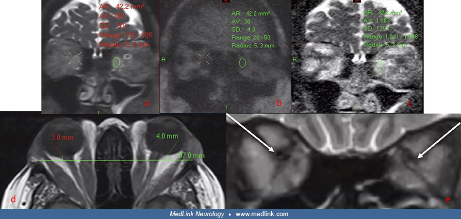
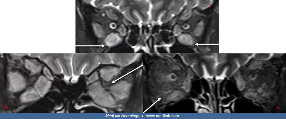
Coronal MRI STIR images: (a) Muscle enlargement with elevated signal intensity in the inferior rectus muscles bilaterally in a patient with thyroid eye disease and no dysthyroid optic neuropathy. (b) Gross enlargement of all extraocular muscles bilaterally in a patient with thyroid eye disease and dysthyroid optic neuropathy. This patient also has apical crowding (arrow) on the left side. (c) Identifying right peri‐muscular fat signal intensity compared to the left orbit, which is less pronounced in a patient with thyroid eye disease. (From: Moledina M, Lee V, Bhatia K, et al. Radiological Activity Score [RAS]-MRI characteristics in dysthyroid optic neuropathy in a multi-ethnic thyroid eye disease population. Clin Endocrinol [Oxf] 2025;103[3]:385-95. Creative Commons Attribution 4.0 International [CC BY 4.0] license, creativecommons.org/licenses/by/4.0.)