Neurobehavioral & Cognitive Disorders
Autism spectrum disorder
Jan. 27, 2026
MedLink, LLC
3525 Del Mar Heights Rd, Ste 304
San Diego, CA 92130-2122
Toll Free (U.S. + Canada): 800-452-2400
US Number: +1-619-640-4660
Support: service@medlink.com
Editor: editor@medlink.com
ISSN: 2831-9125
Toll Free (U.S. + Canada): 800-452-2400
US Number: +1-619-640-4660
Support: service@medlink.com
Editor: editor@medlink.com
ISSN: 2831-9125
Nearly 3,000 illustrations, including video clips of neurologic disorders.
Every article is reviewed by our esteemed Editorial Board for accuracy and currency.
Full spectrum of neurology in 1,200 comprehensive articles.
Listen to MedLink on the go with Audio versions of each article.
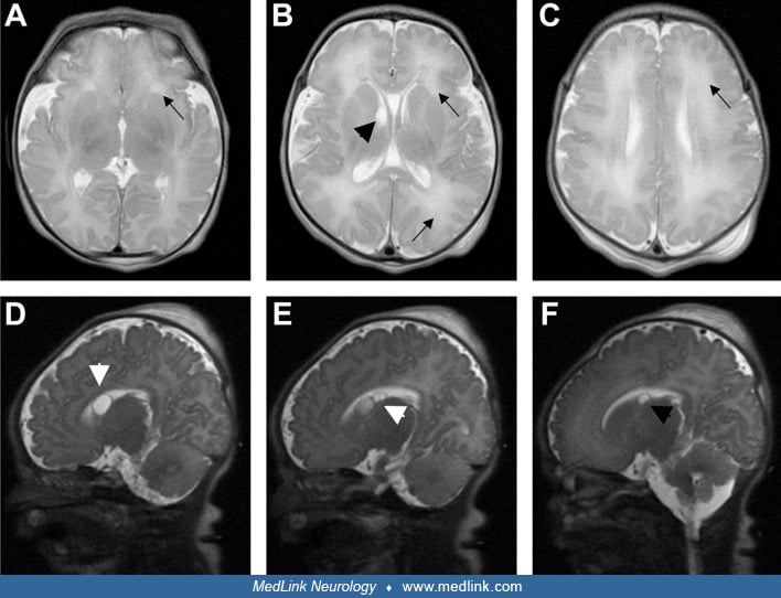
MRI imaging from a critically ill neonate who presented with clinical and electrographic seizures and respiratory failure within 24 hours of life and was subsequently diagnosed with Zellweger syndrome, which was initially identified on newborn metabolic screen for ALD prior to return of any diagnostic testing. (A-C) Axial T2-weighted Propeller magnetic resonance imaging at multiple cerebral levels demonstrates leukodystrophic changes typical of Zellweger disease (black arrows) and one of several bilateral caudothalamic groove cysts (B, large black arrowhead) identified in this individual. (D-F) Sagittal T2 SSFSE magnetic resonance imaging of the same neonate demonstrates multiple bilateral caudothalamic groove cysts (white arrowheads). (Contributed by Dr. Kimberly Chapman.)


