Sleep Disorders
Sleep, stroke, and vascular dementia
Oct. 13, 2025
MedLink, LLC
3525 Del Mar Heights Rd, Ste 304
San Diego, CA 92130-2122
Toll Free (U.S. + Canada): 800-452-2400
US Number: +1-619-640-4660
Support: service@medlink.com
Editor: editor@medlink.com
ISSN: 2831-9125
Toll Free (U.S. + Canada): 800-452-2400
US Number: +1-619-640-4660
Support: service@medlink.com
Editor: editor@medlink.com
ISSN: 2831-9125
Nearly 3,000 illustrations, including video clips of neurologic disorders.
Every article is reviewed by our esteemed Editorial Board for accuracy and currency.
Full spectrum of neurology in 1,200 comprehensive articles.
Listen to MedLink on the go with Audio versions of each article.
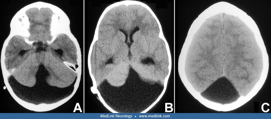
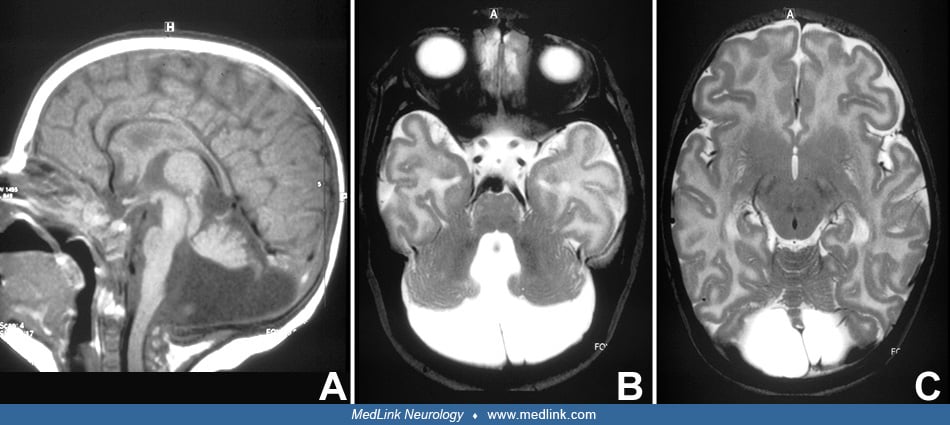
(A) Mid-sagittal view: posterior fossa appears large. Small anterior vermis. Posterior part of corpus callosum is hypoplastic. (B) Axial section through posterior fossa: enlarged 4th ventricle with broad communication to fluid-filled posterior fossa. Cerebellar hemispheres are hypoplastic. (C) Axial section at mid-brain level demonstrating anterior vermis and posterior fossa cyst. (Contributed by Dr. Eugen Boltshauser.)