Sialidosis
Dec. 02, 2025
MedLink, LLC
3525 Del Mar Heights Rd, Ste 304
San Diego, CA 92130-2122
Toll Free (U.S. + Canada): 800-452-2400
US Number: +1-619-640-4660
Support: service@medlink.com
Editor: editor@medlink.com
ISSN: 2831-9125
Toll Free (U.S. + Canada): 800-452-2400
US Number: +1-619-640-4660
Support: service@medlink.com
Editor: editor@medlink.com
ISSN: 2831-9125
Nearly 3,000 illustrations, including video clips of neurologic disorders.
Every article is reviewed by our esteemed Editorial Board for accuracy and currency.
Full spectrum of neurology in 1,200 comprehensive articles.
Listen to MedLink on the go with Audio versions of each article.
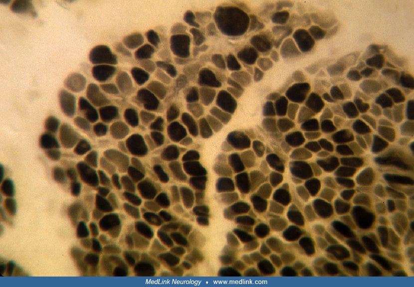
This biopsy is from a 2-month-old term infant with congenital myotonic dystrophy. In this pattern, centronuclear fibers are few, but 2 populations of fibers are seen by size: large (normal sized) fibers and atrophic (more likely hypoplastic) rounded to angular fibers. Although the surface area is dominated by the larger fibers, histograms actually show a predominance of the small fibers. Histochemical stains reveal that the small fibers are nearly exclusively type I and the normal fibers are type II, corresponding to congenital muscle fiber-type disproportion. Modified Gomori trichrome stain. X250. (Contributed by Dr. Harvey Sarnat.)