Neuropharmacology & Neurotherapeutics
Galantamine
Mar. 20, 2021
MedLink, LLC
3525 Del Mar Heights Rd, Ste 304
San Diego, CA 92130-2122
Toll Free (U.S. + Canada): 800-452-2400
US Number: +1-619-640-4660
Support: service@medlink.com
Editor: editor@medlink.com
ISSN: 2831-9125
Toll Free (U.S. + Canada): 800-452-2400
US Number: +1-619-640-4660
Support: service@medlink.com
Editor: editor@medlink.com
ISSN: 2831-9125
Nearly 3,000 illustrations, including video clips of neurologic disorders.
Every article is reviewed by our esteemed Editorial Board for accuracy and currency.
Full spectrum of neurology in 1,200 comprehensive articles.
Listen to MedLink on the go with Audio versions of each article.
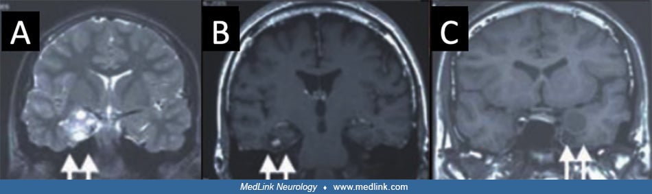
An illustration of the function of coronal MRI slices oblique to the axis of the hippocampus showing right hippocampal sclerosis with volume loss and hypointensity on T1-weighted images (A), FLAIR hyperintensity (B). This is in comparison to another patient with normal hippocampal structure on T2-weighted imaging (C) but with loss of grey-white matter differentiation in the lateral temporal lobe (D). (Image from: Maehara T. Neuroimaging of epilepsy. Neuropathology 2007;27(6):585-93.)