Neurobehavioral & Cognitive Disorders
Aphantasia
Jan. 20, 2026
MedLink, LLC
3525 Del Mar Heights Rd, Ste 304
San Diego, CA 92130-2122
Toll Free (U.S. + Canada): 800-452-2400
US Number: +1-619-640-4660
Support: service@medlink.com
Editor: editor@medlink.com
ISSN: 2831-9125
Toll Free (U.S. + Canada): 800-452-2400
US Number: +1-619-640-4660
Support: service@medlink.com
Editor: editor@medlink.com
ISSN: 2831-9125
Nearly 3,000 illustrations, including video clips of neurologic disorders.
Every article is reviewed by our esteemed Editorial Board for accuracy and currency.
Full spectrum of neurology in 1,200 comprehensive articles.
Listen to MedLink on the go with Audio versions of each article.
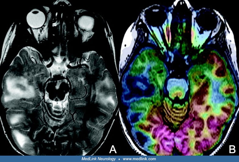
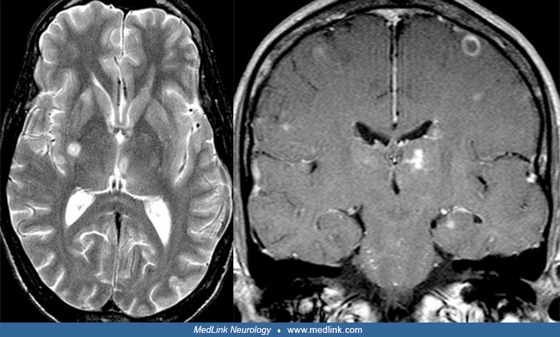
Identification of a hypothalamic hamartoma on various MRI images, all acquired on a 3T scanner. (Top left) T1-weighted sagittal image. (Top right) FLAIR coronal image. (Bottom left) T2-weighted axial image. (Bottom right) postcontrast enhancement T1-weighted axial image. (Contributed by Dr. Noriko Salamon.)