Pyruvate dehydrogenase complex deficiency
May. 28, 2024
MedLink®, LLC
3525 Del Mar Heights Rd, Ste 304
San Diego, CA 92130-2122
Toll Free (U.S. + Canada): 800-452-2400
US Number: +1-619-640-4660
Support: service@medlink.com
Editor: editor@medlink.com
ISSN: 2831-9125
Toll Free (U.S. + Canada): 800-452-2400
US Number: +1-619-640-4660
Support: service@medlink.com
Editor: editor@medlink.com
ISSN: 2831-9125
Nearly 3,000 illustrations, including video clips of neurologic disorders.
Every article is reviewed by our esteemed Editorial Board for accuracy and currency.
Full spectrum of neurology in 1,200 comprehensive articles.
Listen to MedLink on the go with Audio versions of each article.
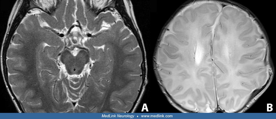
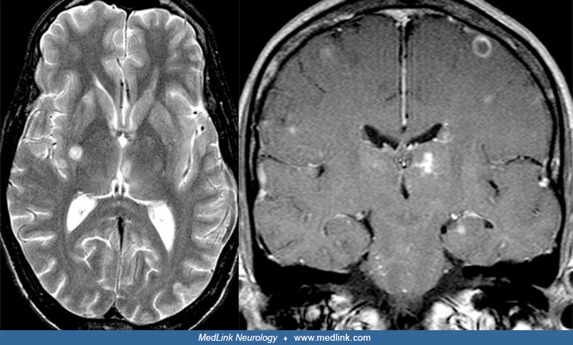
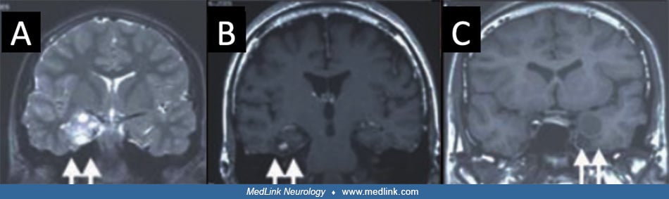
A 42-year-old female with no prior history of seizures presented with status epilepticus thought to be due to antibody negative autoimmune limbic encephalitis. (A) B1500 diffusion weighted imaging shows diffusion restriction in right mesial temporal lobe. (B) Apparent diffusion coefficient (ADC) illustrates acute timing of diffusion restriction. (C) Fluid-attenuated inversion recovery (FLAIR) shows similar right mesial temporal hyperintensity. (Contributed by Dr. Wesley Kerr.)