Movement Disorders
Tourette syndrome
Mar. 01, 2024
MedLink®, LLC
3525 Del Mar Heights Rd, Ste 304
San Diego, CA 92130-2122
Toll Free (U.S. + Canada): 800-452-2400
US Number: +1-619-640-4660
Support: service@medlink.com
Editor: editor@medlink.com
ISSN: 2831-9125
Toll Free (U.S. + Canada): 800-452-2400
US Number: +1-619-640-4660
Support: service@medlink.com
Editor: editor@medlink.com
ISSN: 2831-9125
Nearly 3,000 illustrations, including video clips of neurologic disorders.
Every article is reviewed by our esteemed Editorial Board for accuracy and currency.
Full spectrum of neurology in 1,200 comprehensive articles.
Listen to MedLink on the go with Audio versions of each article.
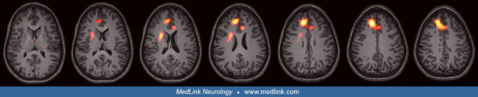
SPECT with left mesial temporal lobe epilepsy (A) and right lateral temporal lobe epilepsy (B). These images are generated by the subtraction of the interictal scan from the ictal scan. Voxels with a significant difference are displayed with yellow being a larger difference and red being less different. (Image from Epilepsy.com.)