Neuro-Oncology
Ependymoma
Jan. 14, 2025
MedLink, LLC
3525 Del Mar Heights Rd, Ste 304
San Diego, CA 92130-2122
Toll Free (U.S. + Canada): 800-452-2400
US Number: +1-619-640-4660
Support: service@medlink.com
Editor: editor@medlink.com
ISSN: 2831-9125
Toll Free (U.S. + Canada): 800-452-2400
US Number: +1-619-640-4660
Support: service@medlink.com
Editor: editor@medlink.com
ISSN: 2831-9125
Nearly 3,000 illustrations, including video clips of neurologic disorders.
Every article is reviewed by our esteemed Editorial Board for accuracy and currency.
Full spectrum of neurology in 1,200 comprehensive articles.
Listen to MedLink on the go with Audio versions of each article.
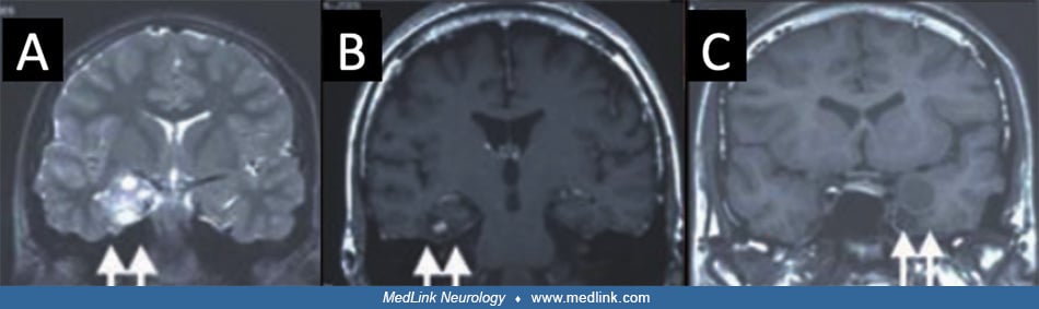
Left occipital hemimegalencephaly suggested by deviation of the sagittal sinus and hypertrophy of the area in a 19-year-old female as seen on brain CT without contrast (A), T1-weighted 1.5T MRI with polymicrogyria (B), and FLAIR white matter hyperintensity (C). (Images from: Feidert A. Hemi-hemimegalencephaly or posterior quadrantic dysplasia, a rare cause of focal epilepsy in an otherwise healthy young woman: a case report. Cureus 2020;12(8):e10002.)