Neuromuscular Disorders
Viral and retroviral myositis
Jun. 16, 2026
MedLink, LLC
3525 Del Mar Heights Rd, Ste 304
San Diego, CA 92130-2122
Toll Free (U.S. + Canada): 800-452-2400
US Number: +1-619-640-4660
Support: service@medlink.com
Editor: editor@medlink.com
ISSN: 2831-9125
Toll Free (U.S. + Canada): 800-452-2400
US Number: +1-619-640-4660
Support: service@medlink.com
Editor: editor@medlink.com
ISSN: 2831-9125
Worddefinition
At vero eos et accusamus et iusto odio dignissimos ducimus qui blanditiis praesentium voluptatum deleniti atque corrupti quos dolores et quas.
Disorders of calcium metabolism, including hyper- or hypofunction of parathyroid hormone, are frequently overlooked causes of muscle dysfunction, and presentation is often non-descript, with mild proximal weakness, muscle pain, and a normal CK. However, when identified and treated, complete resolution of the myopathy can occur, as seen with removal of an adenomatous parathyroid gland for primary hyperparathyroidism or with a subtotal parathyroidectomy for secondary hyperparathyroidism in patients on chronic hemodialysis. There are two mechanisms in which primary hyperparathyroidism can cause weakness: decreased energy production and skeletal muscle catabolism. However, the pathophysiology of weakness in hypoparathyroidism is not well understood.
|
• Primary hyperparathyroidism, secondary hyperparathyroidism (due to renal failure), and osteomalacia may cause myalgia, mild proximal weakness, and normal or slightly elevated CK; the shared features suggest a similar effect on muscle metabolism due to parathyroid hormone excess and vitamin D deficiency. | |
|
• Primary hyperparathyroidism is a common cause of hypercalcemia and hypomagnesia, which can cause a non-necrotizing proximal myopathy. It can also lead to hypophosphatemia, which can rarely result in a necrotizing myopathy associated with rhabdomyolysis. | |
|
• Myopathy (with normal or elevated CK) is rare in primary hypoparathyroidism. The most common muscle manifestation in hypoparathyroidism is due to hypocalcemia, resulting in tetany (hyperexcitability of nerve axons leading to repetitive firing) and subsequent muscle cramps or spasms (carpopedal or laryngeal). | |
|
• Management of myopathies related to parathyroid disorders requires treatment of the primary cause, eg, removal of a hyperfunctioning adenoma in primary hyperparathyroidism; vitamin D and calcium replacement in osteomalacia; removal of hyperfunctioning parathyroid glands and treatment with cholecalciferol (D3) or kidney transplant in secondary hyperparathyroidism due to chronic kidney failure. |
Muscle weakness is common in disorders of calcium and phosphorous homeostasis, including primary and secondary hyperparathyroidism, osteomalacia, hypoparathyroidism, and other abnormalities of bone metabolism. The association of myopathy with hyperparathyroidism and osteomalacia was recognized over a century ago by German physician Karl Hirschberg and German pathologist Friedrich Daniel von Recklinghausen (1833–1910) (57; 136). Vicale reintroduced this association in the modern era, describing three patients with severe proximal weakness, waddling gait, and extensive bone disease (135). Patten and colleagues thought that the clinical features, electromyography, and muscle biopsies from weak patients with hyperparathyroidism reflected a neurogenic disorder (101). However, others disputed this and suspected a myopathic basis for the weakness (79; 71).
In contrast to hyperparathyroidism and osteomalacia, hypoparathyroidism has only rarely been associated with overt clinical myopathy (139; 128; 74; 140). Patients can have elevated serum creatine kinase levels and myopathic features in electrophysiologic or histologic studies, which may be secondary to tetany or seizures resulting from hypocalcemia (59; 137; 122; 71; 01; 110). However, objective weakness is uncommon (71).
Hyperparathyroidism. The myopathy associated with primary hyperparathyroidism or osteomalacia is characterized by the gradual onset of symmetric proximal weakness with atrophy (125; 126; 77; 66; 99; 111). However, the main symptom is often localized bone tenderness or pain (involving the spine, rib cage, pelvis, and shoulder girdle) or generalized aches (28). The combination of proximal muscle weakness and bone or muscle pain may be the presenting complaint in up to 50% of patients with primary hyperparathyroidism (78; 93). Significant leg weakness leads to a waddling gait or even the inability to walk in some patients (92). In children, a Gowers sign may be present (05). The non-necrotizing limb-girdle myopathy is associated with serum hypercalcemia and hypomagnesium in hyperparathyroidism, whereas hypophosphatemia can cause a necrotizing limb-girdle myopathy (69). Patten and colleagues reported several patients with bulbar involvement as evidenced by hoarseness, dysphagia, and fasciculations (101). Rarely, patients have severe neck extensor weakness (11; 116), with “dropped head” syndrome or severe respiratory muscle involvement (45; 98). The basis for the weakness in osteomalacia is not entirely clear. Cramps and paresthesia are reported in approximately 50% of patients (132). Weakness does not correlate closely with calcium, phosphorus, or parathyroid concentrations.
|
• Generalized fatigue | |
|
• Bone tenderness, bone pain, and generalized aches with antalgic gait (which may be severe and may interfere with confrontational muscle strength testing) | |
|
• Cranial nerves normal (except possibly involuntary, fine, tremulous movements of tongue) | |
|
• Myopathy with slight or moderate muscle wasting, symmetric proximal weakness (pelvic girdle more affected than shoulder girdle), neck flexor weakness, and waddling gait | |
|
• Myalgias | |
|
• No fasciculations | |
|
• Reflexes normal or brisk, possibly with spread | |
|
• Sensation normal (although one group of authors report decreased vibratory and pain sensation) | |
|
• Polyarthralgias | |
|
• Bone fractures | |
|
• Hypocalcemia | |
|
• Perioral/acral paresthesias | |
|
• Carpopedal spasm | |
|
• Chvostek sign (ie, ipsilateral facial muscle contraction induced by slight tap of facial nerve anterior to external auditory meatus) | |
|
• Trousseau sign (ie, elicitation of carpopedal spasm and acral paresthesias by compression of upper arm with a tourniquet or blood pressure cuff) | |
|
• Rarely seizures | |
|
• Hypophosphatemia | |
|
• Palpitations | |
|
• Decreased cardiac output | |
|
• Dyspnea/orthopnea | |
|
• Rarely rhabdomyolysis (with/without renal failure) |
Besides proximal weakness and atrophy, examination of patients with hyperparathyroidism often reveals brisk muscle stretch reflexes with flexor plantar responses, and there are rare reports of spasticity and extensor plantar responses (20; 44). In addition, 29% to 57% of patients experience a stocking-glove distribution of reduced pain or vibratory sensation and decreased muscle stretch reflexes suggestive of an underlying peripheral neuropathy (101; 132). Patten and colleagues also described abnormal tongue movements reminiscent of fasciculations (101), but others have not found this feature. Neurobehavioral abnormalities (ie, memory loss, poor concentration, personality changes, inappropriate behavior, anxiety, and hallucinations) can also occur. Rheumatological symptoms such as arthralgias and myalgias can also be seen (87).
Older patients with primary hyperparathyroidism may have significant functional deficits, including slower performance of repeated sit-to-stand and slower gait speed that may affect safety and quality of life (91). Non-neurologic presentations of primary hyperparathyroidism can include pathologic fractures, recurrent renal stones, acute pancreatitis, or solitary bone swelling (93).
Secondary hyperparathyroidism most often occurs in patients with chronic renal failure, who can develop weakness similar to that observed in primary hyperparathyroidism and osteomalacia. Lower extremity weakness predominates early, with eventual progression to involvement of all four limbs. Rare cases of arterial calcification with secondary muscle necrosis and myoglobinuria have been reported in this condition (108).
Vitamin D deficiency has also been associated with muscle symptoms, including myalgia and proximal myopathy (134). This is a significant public health issue, given the high prevalence of vitamin D deficiency; the symptoms often respond favorably to replacement therapy (58). In 25 patients with chronic kidney disease on dialysis, there was a correlation between reduced quadriceps muscle strength and increased fall risk with low levels of 25-hydroxyvitamin D [25(OH)D] levels, but not with levels of parathyroid hormone (15). This was further supported by several meta-analyses. Stockton and colleagues concluded that muscle strength improved if supplemented with 1000 IU of vitamin D in individuals with a baseline serum 25(OH)D level of less than 25 nmol/L (130). In 2014, Beaudart and colleagues also found a small but statistically significant improvement in muscle strength with vitamin D supplementation (10).
Hypoparathyroidism. Before treatment, a large percentage of patients with hypoparathyroidism have muscle-related complaints, such as muscle twitching, muscle weakness, muscle cramping, and muscle spasms (17). However, myopathy secondary to hypoparathyroidism is unusual, although perioral and distal paresthesia and tetany secondary to hypocalcemia do occur. Chvostek sign (ie, ipsilateral facial contraction when tapping the facial nerve at the external auditory meatus) and Trousseau sign (ie, thumb adduction, metacarpophalangeal joint flexion, and interphalangeal joint extension after temporary occlusion of the brachial artery) may be demonstrated in hypocalcemic patients. Severe tetany can cause stridor and apnea due to vocal cord involvement, as well as bronchospasm, diaphragmatic chest wall contraction (23; 81), and occasionally, tetanic seizures (76).
There are only a few reports of mild proximal weakness in patients with hypoparathyroidism (139; 128; 74; 140), and only a small series of cases of myopathy or myalgia with raised serum creatine kinase (CK) associated with hypocalcemia and hypoparathyroidism (56; 131; 27). A non-necrotizing myopathy may be a partial cause of the muscle weakness seen in some patients (33). Back pain and stiffness are often major complaints, and rare cases of hypoparathyroidism have been misdiagnosed as ankylosing spondylitis in part due to paravertebral ligamentous calcification or ossification and the presence of syndesmophytes (68).
A case of idiopathic hypoparathyroidism with proximal myopathy, high CK, low calcium, and EMG evidence of myokymia and neuromyotonia has been reported (142). There are also reports of painless myoglobinuria without objective weakness or tetany (01; 94). Patients with Kearns-Sayre syndrome, a mitochondrial encephalomyopathy due to sporadic large-scale mitochondrial DNA deletions, may also develop endocrinopathies including hypoparathyroidism (114).
Because more extensive blood chemistries are now performed routinely, hyperparathyroidism is diagnosed earlier than in the past, and patients are frequently asymptomatic or only mildly affected (71). In symptomatic patients, medical or surgical (parathyroidectomy) treatment is effective, and improvement of muscle strength is usually noticeable within a few months (101; 71; 95; 96).
Case 1. Primary hyperparathyroidism and primary vitamin D deficiency (Barohn). A 76-year-old man was referred for a 1-year history of progressive weakness. He complained of difficulty climbing stairs, arising from chairs, and lifting objects over his head. In addition, he noted hoarseness of his voice over the past year.
Evaluation by an otolaryngologist was unrevealing. The patient denied weakness in his hands or feet, ptosis, diplopia or blurred vision, dysarthria, dysphagia, difficulty chewing, nasal regurgitation, fluctuation in muscle strength or voice, myalgia, numbness, or paresthesia.
Past medical history was remarkable for prostate cancer, which was treated with radical prostatectomy and bilateral orchiectomies. There was no evidence of tumor recurrence. He also had chronic renal insufficiency that was attributed to hypertension. There was no family history of neuromuscular disorders. He denied alcohol or tobacco use. Medications included propranolol, diltiazem, and aspirin.
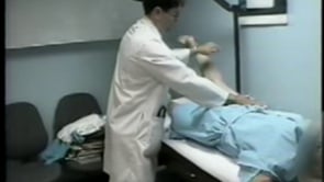
Examination revealed a normal mental status. Extraocular movements were normal. There was no ptosis. Face, jaw, palate, and tongue strength were normal. He had decreased muscle bulk in his shoulder and hip girdles. Fasciculations were not evident. Manual muscle-strength testing demonstrated symmetric proximal upper and lower extremity weakness.
He had the following Medical Research Council grades: orbicularis oculi 5, neck flexors 4, neck extensors 5, shoulder abductors 4, elbow flexors and extensors 4, wrist flexors and extensors 5-, hand intrinsics 5, hip flexors 4-, hip extensors 4-, hip abductors 4-, knee flexors 4, knee extensors 5, ankle dorsiflexors 4+, and plantar flexors 5. Complex motor skills were intact. He had diminished pain and vibratory sensation in his feet to the distal one third of his lower legs bilaterally. His gait was slightly wide-based, but he was able to walk on his heels and toes. Muscle stretch reflexes were 1+ at his biceps, triceps, brachioradialis, and knees, but unobtainable at the ankles.
Nerve conduction studies demonstrated a mild sensorimotor polyneuropathy with unobtainable sural sensory nerve action potentials (SNAPs), borderline amplitudes, and mildly prolonged distal latencies of the median, ulnar, and radial SNAPs. He had low-amplitude peroneal compound motor action potentials (CMAPs), borderline-amplitude median CMAPs, but normal ulnar and posterior tibial CMAPs as well as normal distal motor latencies and conduction velocities. There was no evidence of abnormal decrement or increment on repetitive nerve stimulation. EMG of weak proximal muscles demonstrated normal insertional activity and no abnormal spontaneous activity (ie, fibrillations and fasciculations were not present).
There was a mixture of small and large motor unit potentials, but the overall size on quantitation of 20 motor unit potentials was normal. However, the motor unit potentials did appear to recruit early.
Laboratory evaluation was remarkable for anemia (hematocrit 30%) and chronic renal insufficiency (blood urea nitrogen 32 mg/dL and creatinine 1.9 mg/dL). In addition, he had a low serum albumin level of 2.0 g/dL. Of note, the alkaline phosphatase and electrolytes were normal, including a calcium level of 8.4 mg/dL and phosphate level of 4.3 mg/dL. However, an ionized calcium level (correcting for the hypoalbuminemia) was mildly elevated at 5.52 mg/dL (normal 4.48 to 5.20 mg/dL). Intact parathyroid hormone was elevated at 106.5 pg/mL (normal 10 to 65 pg/dL). His 25-hydroxy vitamin D level was low at 8.6 ng/mL (normal range for the reference lab was 16 to 74 ng/mL), and the 1,25 dihydroxy vitamin D level was not measurable. Other pertinent blood work revealed “normal" serum immunofixation, fasting glucose, creatine kinase, thyroid and liver function tests, erythrocyte sedimentation rate, rheumatoid factor, antinuclear antibody, B12, folate, and CSF studies (protein 32 mg/dL). Acetylcholine receptor antibodies were not present. Nuclear medicine scans demonstrated increased uptake in the left inferior parathyroid gland.
This patient had the unusual combination of both primary hyperparathyroidism and primary vitamin D deficiency. The patient underwent excision of the parathyroid gland and was started on vitamin D replacement. One year after treatment, he had normal strength in his arms but was still hoarse and had mild weakness in his legs.
Case 2. Severe osteomalacia after bariatric surgery (02). A 39-year-old man who had laparoscopic biliopancreatic diversion 10 years before presentation was not compliant with vitamin and mineral supplements for over 9 years, which led to vitamin D deficiency, secondary hyperparathyroidism, myopathy, muscle atrophy, fragility fractures, hypocalcemia, and other electrolyte disturbances.
Approximately 2 years after the surgery, he complained of fatigue and proximal muscle weakness. By 3 years after the surgery, he complained of numbness of his extremities, leg tremors, hand spasms, and progressive limb weakness, causing gait difficulty, for which he started using walking aids. Around that time, he sought medical advice from a general practitioner who provided reassurance and muscle relaxants without further evaluation. His condition worsened until he became completely bedridden 5 years after surgery. He also complained of confusion, memory loss, disturbed sleep, daytime somnolence, headaches, dysphagia with solid food, constipation, urinary incontinence, and bedsores. On evaluation, he looked ill and in pain, with loose hair, stomatitis, glossitis, and general skin dryness. He was alert but confused. Musculoskeletal examination revealed general muscle wasting and left knee and right ankle deformities. Neurologic examination revealed 1/5 power in the legs and 3/5 power in the arms, with intact sensory function. Laboratory studies revealed severe vitamin D deficiency with severe hypocalcemia. Secondary hyperparathyroidism was discovered: calcium 0.92 mmol/L (reference range: 2.1–2.6 mmol/L); phosphorus 0.56 mmol/L (reference range: 0.81–1.58 mmol/L); alkaline phosphatase 1017 IU/L (reference range: 50–136 IU/L); vitamin D3 0.1 ng/mL (reference range: 30–100 ng/mL); parathyroid hormone 59.5 pmol/L (reference range: 1.6–7 pmol/L). X-ray studies revealed multiple fragility fractures.
After 10 months of vitamin D deficiency treatment and correction of his electrolyte imbalance, his memory function and strength improved. He was able to start standing on his legs, move with walking aids, roll over to each side of his bed, feed himself, and open a can.
Case 3. Parathyroid carcinoma with myopathy (31). A 24-year-old woman who presented with proximal weakness was found to have hypercalcemia. Her serum corrected total calcium was 15 mg/dl (reference range: 8.5–10.3 mg/dl), serum phosphate 2.3 mg/dl (reference range: 2.5–4.5 mg/dl), intact PTH 118 pg/ml (reference range: 20–80), vitamin D 15 ng/ml (low), and urinary calcium/urinary creatinine ratio 2.1 (reference range: 0.1–0.2). Her contrast-enhanced neck CT revealed a well-defined mass posterior to the right lobe of the thyroid measuring 2.6 cm × 2.5 cm × 2.9 cm and an enhancing lesion in the right maxillary sinus, measuring 1.9 cm × 2.3 cm in size. She was started on vitamin D supplementation and underwent right lower focal parathyroidectomy, with normalization of her PTH levels following surgery. Surgical pathology revealed an atypical parathyroid adenoma. She was treated with calcium and vitamin D. Her follow-up was initially uneventful.
(Source: Dematapitiya C, Perera C, Pathmanathan S, et al. Parathyroid carcinoma during pregnancy: a novel pathogenic CDC73 mutation - a case report. BMC Endocr Disord 2022;22[1]:259. Creative Commons Attribution 4.0 Internation...
She became pregnant 1 year following her surgery and presented with a rapidly enhancing neck mass of a 1-week duration at 16 weeks’ gestational age. Laboratory studies suggested recurrent primary hyperparathyroidism: serum corrected total calcium 10.8 mg/dl (reference range: 8.5–10.3 mg/dl), serum phosphate 2.0 mg/dl (reference range: 2.5–4.5 mg/dl), vitamin D 45 ng/ml (normal), and intact PTH 540 pg/ml (reference range: 20–80 pg/ml). She had an elevated spot urine calcium to creatinine ratio of 1.5 but no evidence of nephrolithiasis.
Neck ultrasound revealed a well-defined discrete hypoechoic nodule superior to the thyroid isthmus, which was confirmed by noncontrast MRI of the neck. She underwent an uncomplicated second-trimester parathyroid tumor excision, with normalization of postoperative PTH levels. Surgical pathology revealed a parathyroid carcinoma with vascular and capsular invasion. Her genetic studies revealed a novel frameshift mutation of the CDC73 gene. With calcium and vitamin D supplementation, her ionized calcium and PTH levels remained normal throughout the remainder of her pregnancy. She had an uncomplicated cesarean section at 37 weeks.
Case 4. A 38-year-old woman complained of progressive, predominantly proximal weakness and wasting of all four limbs for 3 months. She could not walk without assistance. After admission, she was anxious and developed repeated vomiting, increased thirst, frequent urination, and recurrent colicky upper abdominal pain. She had no family history of endocrine or neoplastic diseases. A small non-tender round nodule measuring approximately 2 × 1.5 cm was noted over the right thyroid lobe. She had wasting of the proximal muscles of all four limbs, especially of the legs and particularly the quadriceps. No fasciculations were noted. Muscle power (MRC scale) was grade 3 proximally and grade 5 distally in all four limbs. Gait was waddling, and Gowers' sign was present. Muscle stretch reflexes were exaggerated, and the plantar reflexes were flexor.
Admission laboratory studies showed severe hypercalcemia with marked elevation of intact parathyroid hormone levels: corrected serum calcium 16 mg/dL (reference range: 8.5–10.5 mg/dL), intact parathyroid hormone (iPTH) 677.3 pg/mL (reference range: 7–53 pg/mL), and inorganic phosphate 2.3 mg/dL (reference range: 2.3–4.7 mg/dL). She was treated with oral and intravenous fluids, injectable proton pump inhibitors, and intravenous zoledronic acid. Her electrocardiogram was normal. After 5 days, serum calcium level dropped to 10 mg/dL, but the iPTH level increased to 1374.2 pg/mL. Ultrasonography of the neck revealed two nodular lesions at the right lobe of the thyroid gland: one was a large (27 × 24 mm) complex nodule in the lower pole and the other was a smaller (17 × 14 mm) nodule in the upper pole. Tc99m sestamibi scan showed increased radiotracer uptake in the area of the right thyroid gland, suggestive of parathyroid adenoma/carcinoma.
A skeletal survey was normal. Renal and thyroid function tests, additional endocrine screening tests (to assess for multiple endocrine neoplasia), serum amylase, creatine kinase, blood sugar, and renal and abdominal imaging revealed no abnormalities. Serum alkaline phosphatase level was slightly increased at 178 U/L (30–120 U/L). Nerve conduction velocities were normal. Electromyography showed full recruitment, mixed short and normal duration, small amplitude polyphasic potentials in different muscles (quadriceps, biceps brachii, etc.), consistent with myopathic potentials.
After resection of the nodules, two biopsied specimens were sent for histopathology: the larger nodule in the lower pole was diagnosed as papillary thyroid carcinoma, and the smaller nodule in the upper pole was diagnosed as parathyroid adenoma. Post-surgery serum calcium and iPTH levels were 9.0 mg/dL and 41.9 pg/mL, respectively. She was again referred for total thyroidectomy.
One month after surgery, her weakness was improved: muscle power was grade 4 proximally and grade 5 distally.
Parathyroid hormone and parathyroid hormone–related protein. Parathyroid hormone (PTH) is an endocrine peptide found exclusively in the parathyroid glands, whereas parathyroid hormone-related protein (PTHrP) has endocrine, paracrine, and autocrine actions and is expressed in a wide range of tissues and organs (129). PTH and PTHrP share the initial 13 amino acid residues at the N-terminus and bind to the same type 1 PTH receptor (PTH1R). The two principal signaling pathways of PTH1R are the cyclic adenosine monophosphate (cAMP) and phosphatidylinositol pathways. An abnormal increase in PTH production can occur in primary and secondary hyperparathyroidism, whereas PTHrP can be produced in large quantities by solid organ malignancies.
(A) Amino acid sequences of the N-terminal region of PTH and PTHrP. The shared sequences with PTH are indicated by dark purple circles on PTHrP. (B) Binding of PTH and PTHrP to a G-protein-coupled receptor. (C) The two principa...
Calcium and phosphorus homeostasis. Calcium and phosphorus homeostasis require a complex interaction of intestinal, renal, hepatic, endocrine, skin, and skeletal functions (71). PTH regulates blood calcium concentration by promoting bone resorption, reducing renal clearance of calcium, and enhancing conversion of 25-hydroxy vitamin D to the active form, 1,25 dihydroxy vitamin D. In addition, intestinal calcium absorption is increased under the influence of vitamin D (112; 29).
There are several forms of vitamin D: (1) vitamin D3 or cholecalciferol, which is derived from the skin; (2) vitamin D2 or ergocalciferol, which is dietary and absorbed through the intestines; and (3) 25-hydroxy vitamin D, which is made in the liver and converted to the more potent metabolite 1,25 dihydroxy vitamin D in the kidney. Increased PTH leads to an increased synthesis of 1,25-dihydroxy vitamin D, hypercalcemia, and hypophosphatemia. Serum phosphorus levels are also determined by diet, intestinal absorption, and renal excretion. Persistently elevated PTH results in resorption of minerals within bone and replacement by fibrous tissue, a condition termed "osteitis fibrosa" or "osteitis fibrosa cystica" in severe forms (71).
Primary and secondary hyperparathyroidism. Primary hyperparathyroidism is most often caused by a single overactive parathyroid gland, usually an adenoma or, rarely (1%), a carcinoma. In 15% of cases, diffuse hyperplasia of all the parathyroid glands is the culprit (105). Rare genetic disorders such as multiple endocrine neoplasia type 1 can also cause primary hyperparathyroidism (51). Effects of elevated parathyroid hormone commonly include hypercalcemia, osteopenia, and nephrolithiasis, although the most common presentation is of asymptomatic laboratory abnormalities.
Secondary hyperparathyroidism is a condition in which a disease outside of the parathyroid glands causes the parathyroid glands to become enlarged and hyperactive. The most common causes of secondary hyperparathyroidism are chronic renal failure or vitamin D deficiency. Patients with chronic kidney disease have a high rate of severe vitamin D deficiency that is exacerbated by the reduced ability to convert 25-hydroxy vitamin D into the active form, 1,25 dihydroxy-vitamin D. The lack of the active form of vitamin D causes decreased intestinal absorption of calcium. In addition, decreased renal phosphorus clearance causes increased serum phosphorous that binds and sequesters calcium. Both lead to hypocalcemia and signal to increase parathyroid hormone secretion. Severe vitamin D deficiency also leads to secondary hyperparathyroidism and osteomalacia. Osteomalacia ("adult rickets") is characterized by defective mineralization of osteoid, the unmineralized bone matrix synthesized by osteoblasts. Other causes of secondary hyperparathyroidism include gastrointestinal bypass surgery and severe celiac or Crohn disease.
In addition to acquired forms, there are hereditary forms of primary hyperparathyroidism (51) and of vitamin D deficiency and osteomalacia (48). An increased incidence of usually asymptomatic secondary hyperparathyroidism associated with vitamin D deficiency has been found in up to 17.5% of patients with myotonic dystrophy type 1, which correlated with the CTG expansion size (97). Only one of 17 patients was symptomatic and had surgery for an adenoma.
Tumor-induced osteomalacia is a rare paraneoplastic syndrome caused by mesenchymal tumors that secrete fibroblast growth factor 23 (FGF23), which acts on the proximal renal tubules to decrease phosphate reabsorption and also decreases the conversion of 25(OH)D to 1,25(OH)D, which further negatively impacts serum phosphate by decreasing phosphate absorption from the intestinal tract (64). Standard treatment for adults is oral phosphate and 1,25(OH)2D. The maximum dosage of oral phosphate may be limited by the development of secondary hyperparathyroidism, which may progress to tertiary hyperparathyroidism if secondary hyperparathyroidism is not adequately addressed.
Hypoparathyroidism. Hypoparathyroidism is seen in various conditions, including complications of surgery (eg, thyroidectomy, parathyroidectomy), hypomagnesemia or hypermagnesemia, irradiation, drugs, sepsis, infiltrative diseases of the parathyroid, and autoimmune, hereditary, or developmental disorders (35). Decreased parathyroid hormone results in diminished synthesis of 1,25-dihydroxy vitamin D, hypocalcemia, and hyperphosphatemia. Osteomalacia can also develop in association with hypoparathyroidism.
Osteomalacia. The accumulation of unmineralized bone matrix leads to rickets in children and osteomalacia in adults. As noted above, osteomalacia can complicate hyperparathyroidism and rarely hypoparathyroidism. Other etiologies of osteomalacia include vitamin D deficiency, inadequate nutrition, phosphate depletion, acidosis, and renal tubular disorders (48; 134). Vitamin D deficiency is common among the elderly who live in northern latitudes and need to cover their skin, and it should be evaluated in older individuals presenting with weakness and premature fatigue (102).
|
Impaired calcium metabolism | ||||
|
• Dietary vitamin D deficiency (nutritional osteomalacia) | ||||
|
- Low dietary intake of vitamin D (118; 62; 47) | ||||
|
- Malabsorption | ||||
|
-- Post-gastrectomy (70) | ||||
|
-- Gluten-sensitive enteropathy (celiac disease) (18) | ||||
|
-- Intestinal, hepatic, biliary, or pancreatic disease | ||||
|
-- Short gut (or short bowel) syndrome (38; 103) | ||||
|
-- Parenteral nutrition | ||||
|
• Impaired vitamin D metabolism | ||||
|
- Inadequate exposure to sunlight (eg, housebound elderly) | ||||
|
-- Enhanced metabolism of vitamin D and 25-hydroxyvitamin D to inactive compounds | ||||
|
-- Anticonvulsants (phenytoin, phenobarbital, primidone) (86; 32; 113; 75) | ||||
|
-- Rifampin | ||||
|
- Impaired formation of 25-hydroxyvitamin D | ||||
|
-- Liver disease | ||||
|
-- Isoniazid | ||||
|
- Impaired formation of 1,25-dihydroxyvitamin D | ||||
|
-- Renal failure | ||||
|
-- Renal tubular disorders (82) | ||||
|
-- Hypoparathyroidism | ||||
|
-- Pseudohypoparathyroidism | ||||
|
-- Drugs (eg, ketoconazole, isoniazid) | ||||
|
- Inherited and other rare conditions | ||||
|
Impaired phosphate metabolism | ||||
|
• Treatment with phosphate binders (eg, aluminum hydroxide in patients with renal failure) (07; 61) | ||||
|
• Fanconi syndrome (82) | ||||
|
• Inherited (119; 124; 109; 141) | ||||
|
• Other rare conditions (106) | ||||
|
Defective bone matrix (toxic effects on bone mineralization) | ||||
|
• Aluminum (07; 61; 19) | ||||
|
• Bisphosphonates | ||||
|
• Fluoride | ||||
|
• Vitamin A (excess or deficiency) (retinoic acid, which the body makes from vitamin A, stimulates osteoclasts but suppresses osteoblasts) | ||||
Myopathy in primary hyperparathyroidism and osteomalacia. There are two mechanisms causing the weakness and fatigability seen in primary hyperparathyroidism and osteomalacia. Elevated levels of parathyroid hormone (possibly related to the accompanying hypophosphatemia) are associated with impaired energy production, transfer, and utilization (06; 71; 129), as well as enhanced muscle proteolysis (42).
The first mechanism, energy metabolism disorder, occurs because parathyroid hormone interferes with the oxidation of long-chain fatty acids by blocking free carnitine from being transformed into acylcarnitine through the inhibition of carnitine palmitoyltransferase activity. Thus, the acylcarnitine supply in the mitochondria is decreased and energy production begins the fail (127).
Excess PTH and PTHrP can result in protein-energy wasting, malnutrition, and cachexia (129). PTH and PTHrP can stimulate the expression of thermogenic genes, causing adipose tissue browning by binding to PTH1R and activating cyclic adenosine monophosphate (cAMP)-dependent protein kinase A in white adipose tissue. This results in an increase in resting energy expenditure, loss of muscle and fat mass, and weight loss.
Abbreviations: PTH, parathyroid hormone; PTH1R, type 1 PTH receptor; PTHrP, parathyroid hormone-related protein.
(Source: Srisuwarn P, Disthabanchong S. Role of parathyroid hormone and parathyroid hormone-related ...
The second mechanism is through the catabolism of skeletal muscle, which leads to atrophy. This may be the result of cyclic AMP (cAMP) activation of calcium channels, initiated by parathyroid hormone, raising the intracellular calcium to a level sufficient to increase activation of intracellular proteases, thus tipping the molecular homeostatic balance in favor of muscle degradation (08).
In addition, parathyroid hormone may diminish the sensitivity of contractile myofibrillary proteins to calcium and activate a cytoplasmic protease, thus impairing the bioenergetics of muscle (133). Interestingly, calcium and phosphorus levels do not correlate well with the clinical severity of muscle weakness (37; 126; 101).
Vitamin D may also play a role in muscle weakness and has been shown to have a direct effect on muscle: it accelerates amino acid incorporation into muscle proteins (14; 71) and increases the uptake of calcium by sarcoplasmic reticulum and mitochondria (26; 104). In experimental animals with vitamin D deficiency, excitation-contraction coupling is deranged, calcium uptake and storage capacity are impaired, myofibrillar ATPase activity is depressed, and protein synthesis is decreased (13). Vitamin D receptors have been demonstrated in adult mouse skeletal muscles with higher concentrations in precursor cells and younger animals (46). Other in vitro studies demonstrated that 1,25(OH)2D3 increases myocyte precursor differentiation, and vitamin D deficiency accelerates muscle degradation through the ubiquitin pathway (43; 12).
Myopathy in hypoparathyroidism and pseudohypoparathyroidism. The etiology of the rare myopathy associated with hypoparathyroidism is not clear. Elevated serum creatine kinase and mild nonspecific histologic abnormalities on muscle biopsy are generally considered secondary to muscle damage from hypocalcemia, with more severe abnormalities related to the degree and duration of the hypocalcemia (27). Decreased serum calcium concentration causes a shift in the cellular activation potential toward the resting potential (16; 39; 39; 01). Therefore, less current is required to elicit an action potential. Tetany may manifest when plasma calcium plummets to about 6 mg/dL (50). By the same process, central nervous system neuronal hyperexcitability can result in seizures.
The reduced peripheral response to parathyroid hormone in pseudohypoparathyroidism is best described in families with a mutation in the GNAS1 gene coding for the alpha subunit of stimulatory G protein, which couples with the parathyroid hormone receptor to stimulate adenylyl cyclase (09).
Osteomalacia most often affects the elderly. Muscle weakness is common in osteomalacia, occurring in as many as 72% of patients in some series (126). Weakness develops in only 2% to 10% of patients with hyperparathyroidism alone (126; 101; 77). Rarely is overt myopathy evident in hypoparathyroidism (71).
Because better techniques are available for the early diagnosis and treatment of hyperparathyroidism and osteomalacia (53), neuromuscular complications in more recent series are milder (132) than those reported in earlier series (135; 126; 101; 85).
A prospective study of age-related muscle loss in men ages 60 to 85 showed that, in addition to low leisure physical activity, type 2 diabetes, and low testosterone, low parathyroid hormone serum concentration was associated with greater age-related acceleration of muscle loss (107).
Certain populations are clearly at a higher risk of developing hypo- or hyperparathyroid complications. These include people who underwent thyroidectomy because the parathyroids are adherent to the thyroid gland and, therefore, at risk for accidental excision (leading to hypoparathyroidism), or family members of people with multiple endocrine neoplasia, who are at increased risk for the disease themselves. Laboratory surveillance (ie, checking serum calcium levels) of such individuals mitigates the risk of developing sequelae.
No information is available on the prevention of myopathy secondary to hypoparathyroidism.
Osteomalacia is often unrecognized and allowed to progress, with patients being given incorrect and misleading diagnoses (eg, fibromyalgia, psychoneurosis, etc.). The differential diagnosis is summarized in the following table based on initial misdiagnoses in reported cases.
|
• Osteoporosis | |
|
• Degenerative joint disease | |
|
• Rheumatoid arthritis | |
|
• Ankylosing spondylitis | |
|
• Diffuse idiopathic skeletal hyperostosis (DISH) | |
|
• Paget disease | |
|
• Metastatic bone disease (eg, secondary to some radiologic changes) | |
|
• Multiple myeloma | |
|
• Polymyositis | |
|
• Motor neuron disease (eg, weakness, wasting, and brisk reflexes) | |
|
• Muscular dystrophy | |
|
• Polymyalgia rheumatica (eg, shoulder/hip girdle pain and "weakness") | |
|
• Fibromyalgia | |
|
• Psychoneurotic disorders (eg, diffuse pain and fatigue with little clinical) |
The pattern of weakness (ie, proximal greater than distal weakness and legs worse than arms) is not specific for parathyroid-related myopathies and can be observed in other endocrine disorders (eg, hyperthyroidism, hypothyroidism, Cushing syndrome) and with administration or surreptitious use of some hormone medications (eg, levothyroxine, cortisone, prednisone). History of corticosteroid use, thyroid function tests, electrolytes, and cortisol levels can help in distinguishing these endocrine myopathies. Polymyositis and dermatomyositis are associated with proximal limb weakness but, in contrast to parathyroid myopathies, serum creatine kinase levels are usually elevated, EMG demonstrates increased insertional and spontaneous activity (fibrillation potentials, complex repetitive discharges), and muscle biopsies reveal inflammation. Less common causes of proximal weakness in older individuals include limb-girdle muscular dystrophies, Lambert-Eaton syndrome, and myasthenia gravis. These disorders should readily be distinguished by clinical and family history, neuromuscular examination, electrophysiologic testing, muscle histology, and ancillary tests (eg, edrophonium test, acetylcholine receptor antibody titer, P/Q voltage-gated calcium channel antibody titers, and genetic testing).
Finally, there are a few cases of hyperparathyroidism mimicking amyotrophic lateral sclerosis, characterized by spasticity, hyperreflexia, and extensor plantar responses in combination with lower motor neuron weakness (101; 20; 44). Some of these patients improved following resection of parathyroid adenomas. However, with appropriate clinical evaluation these patients never would have fulfilled criteria for amyotrophic lateral sclerosis (63). The presence of hyperparathyroidism in patients with amyotrophic lateral sclerosis appears to be coincidental, and patients do not improve following surgical resection. In one study, five patients who met El Escorial criteria for amyotrophic lateral sclerosis were found to have hyperparathyroidism secondary to parathyroid adenomas (63). Resection of the adenomas failed to alter the fatal course of amyotrophic lateral sclerosis in these patients. The previously reported patients who improved with surgical resection of the parathyroid adenomas most likely had the associated myopathy, not amyotrophic lateral sclerosis.
Blood and urine studies. In osteomalacia, serum calcium and phosphate may be decreased or normal, serum alkaline phosphatase increased or normal, serum parathyroid hormone increased, serum phosphate variably low (depending on the degree of secondary hyperparathyroidism), plasma 25-hydroxy vitamin D decreased, and plasma 1,25-dihydroxy-vitamin D decreased or normal. Elevated serum alkaline phosphatase levels are present in 80% to 90% of cases and can serve as a useful screening tool for osteomalacia (115). Creatine kinase levels are typically normal but may be slightly increased in a minority of patients. Other blood and urine studies that may be helpful and that should be considered include serum creatinine, hemoglobin, mean corpuscular volume, urinalysis, and 24-hour urine calcium and phosphorus concentrations. Urinary excretion of calcium is low (except in cases secondary to renal tubular acidosis), whereas excretion of phosphate is high.
In hyperparathyroidism, serum calcium levels are usually elevated and serum phosphate levels are low, whereas urinary excretion of calcium is low and urinary excretion of phosphate is high. In patients with concurrent hypoalbuminemia, serum calcium levels may be normal, and in these cases, ionized calcium should be measured and is typically elevated. Increased urinary excretion of cyclic adenosine monophosphate in the presence of hypercalcemia is indicative of hyperparathyroidism. In primary hyperparathyroidism, serum parathyroid hormone levels are elevated. In hyperparathyroidism secondary to renal failure, 1,25-dihydroxy vitamin D levels are low. Noninvasive imaging techniques, such as ultrasound, thallium/technetium scintigraphy, computed tomography, and magnetic resonance imaging, may be useful in localizing abnormal parathyroid glands (95).
Clinical guidelines are now available for the diagnosis and management of tumor-induced osteomalacia (64).
Hypoparathyroidism is associated with low serum parathyroid hormone and calcium levels, low serum magnesium, and high serum phosphate levels. Although serum creatine kinase is usually normal in hyperparathyroidism, in hypoparathyroidism creatine kinase levels may be elevated. Muscle biopsies may reveal nonspecific myopathic features that may reflect muscle damage secondary to episodes of tetany (71). Decreased glycogen phosphorylase activity of muscle biopsy specimens has also been described (133). Electrocardiogram should be routinely performed to monitor for prolonged QT intervals. Head computed tomography may incidentally show basal ganglia calcifications.
Blood studies in various metabolic bone diseases are summarized in the following table.
|
Condition |
Serum calcium |
Serum phosphate |
Serum alkaline phosphate |
Serum parathyroid hormone |
Plasma 25-hydroxy-vitamin D |
Plasma 1,25-dihydroxy-vitamin D |
|
Vitamin D deficiency |
↓↔ |
↓↔ |
↑↔ |
↑ |
↓ |
↓↔ |
|
Tumor-induced osteomalacia |
↓↔ |
↓ |
↑ |
↑↔ |
↔ |
↓↔ |
|
2° Hyperparathyroidism (chronic kidney disease) |
↓ |
↑ |
↑ |
↑ |
↔ |
↓ |
|
Hypoparathyroidism |
↓ |
↑ |
↔ |
↓ |
↔ |
↓ |
|
Pseudohypoparathyroidism |
↓ |
↑ |
↔ |
↑ |
↔ |
↓ |
|
1° Hyperparathyroidism |
↑ |
↓ |
↑? |
↑ |
↔ |
↔ |
|
Osteoporosis |
↔ |
↔ |
↔ |
↔ |
↔ |
↔ |
|
Paget disease |
↔ |
↔ |
↑ |
↔ |
↔ |
↔ |
Electromyography. In patients with weakness secondary to either hyperparathyroidism or osteomalacia, electromyography may either be normal (20%) or it may show nonspecific myopathic changes (approximately 80%), with brief duration, polyphasic motor unit action potentials of normal or small amplitude with early recruitment (125; 126; 37; 101; 123; 62; 78). There are only rare reports of neurogenic features on EMG, such as fibrillation and fasciculation potentials, large polyphasic motor units, and decreased recruitment (101; 44). Rare reports of myotonic discharges on EMG have been reported (52). Ljunghall and colleagues reported increased jitter on single-fiber EMG but no significant blocking (83). Nerve conduction velocities are typically normal, but slight slowing has been noted in some patients, possibly from other causes (eg, a superimposed axonal sensorimotor neuropathy) (132).
Muscle biopsy. Muscle biopsies usually demonstrate nonspecific myopathic features with nonspecific atrophy or type II fiber atrophy and, in some cases, fatty infiltration, interstitial fibrosis, variation in fiber size, and loss of myofibrils. However, no necrosis of muscle fibers or phagocytosis of fibers has been reported (78).
Roentgenography. In osteomalacia, roentgenography usually demonstrates a decrease in bone density associated with loss of trabeculae, blurring of trabecular margins, and variably thinned cortices (48). X-ray changes in osteomalacia are summarized in the following table.
|
• Generalized demineralization/osteopenia (28) | ||
|
• Radiolucent bands perpendicular to bone surfaces ("pseudofractures" or looser zones) (21; 28; 109) | ||
|
- Pubic rami | ||
|
- Ribs | ||
|
• Rachitic changes in extremity bones (eg, upper humerus, tibia, metatarsals) | ||
|
• Biconcave ("codfish tail") vertebral bodies (28) | ||
|
• Signs of secondary hyperparathyroidism | ||
|
- Phalangeal tuft resorption | ||
|
- Erosion of shafts of metacarpals/phalanges | ||
|
• Fractures | ||
Bone biopsy. In osteomalacic myopathy, a low 25-hydroxy-vitamin D level and characteristic radiographic findings may be adequate for diagnosis. However, a bone biopsy is sometimes recommended to confirm the diagnosis, especially if neither the 25-hydroxy-vitamin D level nor radiographic findings are diagnostic. The bone biopsy is performed after double tetracycline labeling of bone, usually with demeclocycline. Demeclocycline (300 mg orally twice a day) is given for 3 days, then no drug for 14 days, and then demeclocycline is again given for 3 more days prior to a biopsy of the iliac crest.
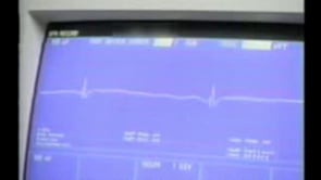
Electrocardiogram (EKG). The main EKG abnormality seen with hypercalcemia (as in hyperparathyroidism) is shortening of the QT interval. Osborn waves (J waves) may be seen in severe hypercalcemia; J waves are a positive deflection in precordial and true limb leads at the J point (where the QRS complex joins the ST segment, the approximate end of depolarization and the beginning of repolarization). Ventricular irritability and ventricular fibrillation have been reported with extreme hypercalcemia.
Hypocalcemia (as in hypoparathyroidism) causes QTc prolongation primarily by prolonging the ST segment (41; 04; 121).
A 12-lead electrocardiogram of a 45-year-old woman showing a QTc interval of 486 ms (suggestive of borderline prolongation) and a pattern of acute anteroseptal injury. She presented in a wheelchair with complaints of generalize...
The T wave is typically unchanged. Dysrhythmias are uncommon, although atrial fibrillation has been reported. Torsades de pointes may occur but is much less common than with hypokalemia or hypomagnesemia.
Parathyroidectomy is the treatment of choice for symptomatic patients with primary hyperparathyroidism (95). In patients with adenoma, the affected gland is removed, although additional glands may be biopsied. Patients with hyperplasia of all four glands generally have subtotal (3.5 glands) parathyroidectomies. Medical management of primary hyperparathyroidism is reserved for asymptomatic patients or those with significant perioperative risk (96). Patients with symptomatic hyperparathyroidism who choose not to have surgery or who are poor surgical candidates can be managed with bisphosphonates and cinacalcet (a calcimimetic that mimics the action of calcium on tissues by allosteric activation of the calcium-sensing receptor) (116).
Patients with secondary hyperparathyroidism usually improve with vitamin D and calcium replacement and lowering of phosphate levels or, if they are in end-stage renal failure, with renal transplantation (133; 84). Cinacalcet can also be added if parathyroid hormone levels remain elevated.
Occasionally, parathyroidectomy is performed in patients with secondary hyperparathyroidism: the three main types of surgery for secondary hyperparathyroidism are subtotal parathyroidectomy (ie, removal of three-and-a-half of the parathyroids), four gland excision and autotransplantation (ie, removing all four parathyroid glands and placing a piece of a parathyroid in the forearm), and PTH-guided parathyroidectomy (ie, removing enough parathyroid tissue to bring the intraoperative PTH levels to between 200 and 300).
In one study, the outcome was similar between medical versus surgical treatment for patients with severe secondary hyperparathyroidism (90); 25% (four of 16 patients) had improvement in their muscle weakness with a subtotal parathyroidectomy whereas 40% (two of five patients) improved with a total parathyroidectomy.
The myopathy associated with osteomalacia responds well to vitamin D and calcium replacement and to treatment of the underlying responsible condition (125; 126; 85; 120; 123; 62; 48; 115). Dietary vitamin D deficiency may be treated with 5000 IU vitamin D daily or 50,000 IU vitamin D weekly for several weeks until bones heal, followed by long-term treatment with 400 to 2000 IU daily. Many authors also recommend calcium supplements (eg, 500 mg elemental calcium twice daily), particularly if dietary calcium intake is (or is likely to be) low. Serum alkaline phosphatase levels will initially increase with treatment and then normalize as bones heal.
Malabsorption of vitamin D may require treatment with high doses of vitamin D, eg, 50,000 IU weekly or from 5000 to 50,000 IU daily, with the dose adjusted to maintain normal serum levels of 25-hydroxy-vitamin D. Calcium supplementation (1.0 to 1.5 gm/day of elemental calcium in two to three divided doses) and treatment of the underlying gastrointestinal disorder are also indicated. Vitamin D dosing will need to be adjusted based on the status of the underlying disease that is causing malabsorption, both during and after treatment. If a patient fails to respond to oral vitamin D administration, alternatives include 10,000 IU vitamin D2 intramuscularly daily or calcitriol 0.5 to 1.0 µg daily. Serum and 24-hour urine calcium levels and serum 25-hydroxy-vitamin D levels should be monitored every 3 to 6 months to avoid hypercalcemia. Patients with gluten-sensitive enteropathy may not respond to vitamin D in standard doses (even when administered parenterally) until a gluten-free diet is instituted.
Patients with either vitamin D–resistant hypophosphatemic rickets or tumor-induced osteomalacia (oncogenic osteomalacia) require treatment with phosphate supplementation (1 to 2 gm/day of oral neutral phosphate) and calcitriol until serum phosphate and alkaline phosphatase concentrations normalize.
Patients with anticonvulsant-induced osteomalacia (or rifampin-induced osteomalacia, eg, in patients treated for tuberculosis) can initially be treated with 4000 IU vitamin D daily, but some patients may require up to 100,000 IU vitamin D per meter squared of body surface area per week. If possible, anticonvulsants should be changed to those less likely to be associated with osteomalacia (phenytoin and barbiturates are most strongly associated with osteomalacia).
The rare myopathy associated with hypoparathyroidism also improves following correction of hypocalcemia and hyperphosphatemia with vitamin D and calcium (139; 140).
Emergency treatment of hypocalcemic tetany requires intravenous infusion of calcium 15 to 20 milliequivalents per kilogram over 4 to 6 hours. When seizures occur, calcium can be given as a slow intravenous push; 10 to 20 mL of 10% calcium gluconate diluted in 50 to 100 mL dextrose or normal saline intravenously over 10 minutes is recommended; however, cardiac and blood pressure monitoring would be necessary (133; 22). The target serum calcium should be between 8 and 9 mg/dL (34), with further titration based on clinical improvement. Concurrent hypomagnesemia must also be addressed with exogenous supplementation. Should stridor be present, close respiratory monitoring is mandatory, with consideration for emergency tracheostomy (a kit should be brought to the bedside) if obstruction occurs. Chronic management with vitamin D2 (ergocalciferol, dose: 25,000 to 150,000 units daily) should begin in conjunction with oral calcium repletion when the patient can safely take oral medication. In some patients who do not respond to vitamin D2, 1,25(OH)2D3 or 1-OH-D3 may be tried (at a higher cost) to circumvent impaired vitamin D activation (133).
Over 80% of 103 consecutive patients with primary hyperparathyroidism who underwent parathyroidectomy improved after surgery (24). In another large series, all 42 patients over the age of 60 benefited from surgery (30). Fifteen of 17 patients with muscle weakness and 14 of 15 patients with premature fatigue reported significant improvement postoperatively (30). Quantitative muscle testing demonstrated mild improvement (3% to 33%) in isokinetic strength or torque in knee extensors and flexors following surgery (54; 138; 65). The increase in isokinetic strength was noted at higher angular velocities, suggesting that therapy had affected predominantly type 2 (fast-twitch) muscle fibers, which, as noted earlier, are selectively involved in the myopathy associated with hyperparathyroidism. Increases in maximal expiratory pressures have also been reported 6 to 12 months postoperatively (72). In one study, postoperative improvement in strength correlated with higher preoperative calcium levels and worse muscle strength (65), but others have not observed such a correlation (54; 138; 72).
Although similar quantitative data are not available for patients with secondary hyperparathyroidism and osteomalacia, subjective and objective improvement in muscle strength also occurs in most patients with secondary hyperparathyroidism and osteomalacia following treatment of the underlying condition (36; 55; 85; 80; 71; 115). In one center's 10-year experience in subtotal parathyroidectomy of hemodialysis patients, myopathic symptoms were present in 89% of patients before surgery and in only 15% of patients after surgery (67).
The rare myopathy associated with hypoparathyroidism improves following correction of the hypocalcemia and hyperphosphatemia with administration of vitamin D and calcium (139; 140).
Primary hyperparathyroidism is rare during pregnancy (51). However, women of childbearing age account for 5% to 25% of patients with primary hyperparathyroidism (73). The most serious complications are neonatal death, stillbirth, and miscarriage.
In a review of 73 reported cases of pregnant patients with primary hyperparathyroidism, 45 patients (62%) had detailed clinical histories, and of these, 22% had weakness with premature fatigue or myalgias (73). Twenty-three patients underwent surgery during pregnancy, and 18 of them gave birth to healthy children. Fifty women with a total of 79 pregnancies did not undergo surgery and bore 35 healthy children; complications arose in 40 births. Perinatal death occurred in 9% of the operated group and 15% of the nonoperated group. To reduce the risk of complications in both the mother and the child, the authors recommended surgery in the second trimester of pregnancy after the fetal organs have developed and the risk of postoperative miscarriage is less (73).
Primary hyperparathyroidism is uncommon in children. Of 1000 patients who underwent parathyroidectomy over a 15-year period, 2.1% were 20 years old or younger (100). The triad of fatigue, weakness, and/or depression was about twice as common in adolescents (67%). Renal stones (52%) and hypercalcemic crisis (5%) were also more common in children, whereas osteopenia and osteoporosis, common in the adults, were not diagnosed in any of the adolescents. Children had higher presenting serum calcium levels and lower parathyroid hormone concentration. Like adults, 86% of children had single gland disease, and 95% were cured after the first operation.
The risk of complications with anesthesia and surgery in patients with myopathies related to hyperparathyroidism is considered low, with mortality less than 2% and morbidity less than 4% (24; 95). Although there is general concern for cardiac dysrhythmias in patients with hypercalcemia, one series found no increase in intraoperative ventricular dysrhythmias in patients with calcium levels of 11.6 mg/dL (49). No particular anesthetic or anesthesia technique is known to produce a better outcome in patients undergoing surgery for hyperparathyroidism. Particular consideration should, however, be given when using succinylcholine or atracurium because sensitivity to these substances has been reported in patients with hyperparathyroidism (03). This is especially pertinent when the surgeon wants to preserve the ability to test the recurrent laryngeal nerve (not routinely done by most surgeons) during surgery.
All contributors' financial relationships have been reviewed and mitigated to ensure that this and every other article is free from commercial bias.

Douglas J Lanska MD MS MSPH
Dr. Lanska of the University of Wisconsin School of Medicine and Public Health has no relevant financial relationships to disclose.
See ProfileNearly 3,000 illustrations, including video clips of neurologic disorders.
Every article is reviewed by our esteemed Editorial Board for accuracy and currency.
Full spectrum of neurology in 1,200 comprehensive articles.
Listen to MedLink on the go with Audio versions of each article.
MedLink, LLC
3525 Del Mar Heights Rd, Ste 304
San Diego, CA 92130-2122
Toll Free (U.S. + Canada): 800-452-2400
US Number: +1-619-640-4660
Support: service@medlink.com
Editor: editor@medlink.com
ISSN: 2831-9125
Neuromuscular Disorders
Jun. 16, 2026
Neuromuscular Disorders
May. 27, 2026
Neuromuscular Disorders
May. 21, 2026
Developmental Malformations
May. 08, 2026
Neuromuscular Disorders
Apr. 23, 2026
Neuromuscular Disorders
Apr. 23, 2026
Neuromuscular Disorders
Apr. 23, 2026
Neuropharmacology & Neurotherapeutics
Apr. 23, 2026