


Epilepsy & Seizures
Developmental and epileptic encephalopathies
Mar. 31, 2025
MedLink, LLC
3525 Del Mar Heights Rd, Ste 304
San Diego, CA 92130-2122
Toll Free (U.S. + Canada): 800-452-2400
US Number: +1-619-640-4660
Support: service@medlink.com
Editor: editor@medlink.com
ISSN: 2831-9125
Toll Free (U.S. + Canada): 800-452-2400
US Number: +1-619-640-4660
Support: service@medlink.com
Editor: editor@medlink.com
ISSN: 2831-9125
Nearly 3,000 illustrations, including video clips of neurologic disorders.
Every article is reviewed by our esteemed Editorial Board for accuracy and currency.
Full spectrum of neurology in 1,200 comprehensive articles.
Listen to MedLink on the go with Audio versions of each article.
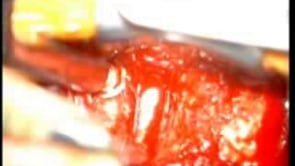
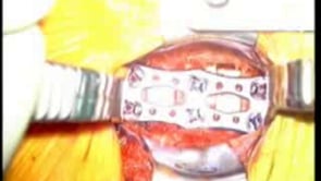
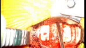
Exposure of anterior cervical microdiscectomy, allograft fusion, and plate fixation for spondylosis. After disc, cartilage, posterior longitudinal ligament, and spur removal, the ventral dural covering the spinal cord at C5-6 is visually inspected to confirm return to normal anatomic position. A nerve hook is passed out laterally through each neural foramen to ensure decompression of the exiting C6 nerve roots. (Left) rostral field; (right) caudal field; (superior) left paracervical retractor blade at longus colli muscle, retracting the esophagus; (inferior) right paracervical retractor blade at longus colli muscle, retracting right carotid sheath. (Contributed by Dr. Saul Schwarz.)