General Neurology
Thoracic outlet syndromes
Nov. 06, 2023
MedLink®, LLC
3525 Del Mar Heights Rd, Ste 304
San Diego, CA 92130-2122
Toll Free (U.S. + Canada): 800-452-2400
US Number: +1-619-640-4660
Support: service@medlink.com
Editor: editor@medlink.com
ISSN: 2831-9125
Toll Free (U.S. + Canada): 800-452-2400
US Number: +1-619-640-4660
Support: service@medlink.com
Editor: editor@medlink.com
ISSN: 2831-9125
Nearly 3,000 illustrations, including video clips of neurologic disorders.
Every article is reviewed by our esteemed Editorial Board for accuracy and currency.
Full spectrum of neurology in 1,200 comprehensive articles.
Listen to MedLink on the go with Audio versions of each article.
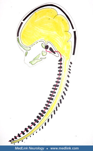
The central nervous system (yellow) and the surrounding axial skeleton in a human fetus (17 weeks' gestation). The notochord (red) is surrounded by the spine, the basilar part of the occipital bone, and the postsphenoid part of the sphenoid body (black). The axial craniofacial skeleton (green) and the thecal skeleton (black). The sella turcica and pituitary gland are located where the black and yellow skeleton fuse in the postsphenoid part of the sphenoid body. The rostral end of the notochord appears close to the sella turcica. (Reproduced with permission. Acta Odontol Scand 1995;53:135-43).