Sleep Disorders
Drug-induced insomnia and excessive daytime sleepiness
Jul. 19, 2021
MedLink, LLC
3525 Del Mar Heights Rd, Ste 304
San Diego, CA 92130-2122
Toll Free (U.S. + Canada): 800-452-2400
US Number: +1-619-640-4660
Support: service@medlink.com
Editor: editor@medlink.com
ISSN: 2831-9125
Toll Free (U.S. + Canada): 800-452-2400
US Number: +1-619-640-4660
Support: service@medlink.com
Editor: editor@medlink.com
ISSN: 2831-9125
Nearly 3,000 illustrations, including video clips of neurologic disorders.
Every article is reviewed by our esteemed Editorial Board for accuracy and currency.
Full spectrum of neurology in 1,200 comprehensive articles.
Listen to MedLink on the go with Audio versions of each article.
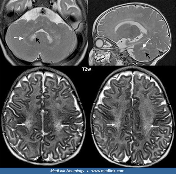
Cerebellar changes in an early infantile patient during the disease course (onset 4 months). The T2-weighted axial MRI at age 5.5 months shows hyperintensity in the dentate nucleus (a), which had disappeared 8 months later (b). (Source: Krieg SI, Krägeloh-Mann I, Groeschel S, et al. Natural history of Krabbe disease: a nationwide study in Germany using clinical and MRI data. Orphanet J Rare Dis 2020;15[1]:243. Creative Commons Attribution 4.0 International [CC BY 4.0] license, creativecommons.org/licenses/by/4.0.)