Sleep Disorders
Narcolepsy
Sep. 02, 2025
MedLink, LLC
3525 Del Mar Heights Rd, Ste 304
San Diego, CA 92130-2122
Toll Free (U.S. + Canada): 800-452-2400
US Number: +1-619-640-4660
Support: service@medlink.com
Editor: editor@medlink.com
ISSN: 2831-9125
Toll Free (U.S. + Canada): 800-452-2400
US Number: +1-619-640-4660
Support: service@medlink.com
Editor: editor@medlink.com
ISSN: 2831-9125
Nearly 3,000 illustrations, including video clips of neurologic disorders.
Every article is reviewed by our esteemed Editorial Board for accuracy and currency.
Full spectrum of neurology in 1,200 comprehensive articles.
Listen to MedLink on the go with Audio versions of each article.
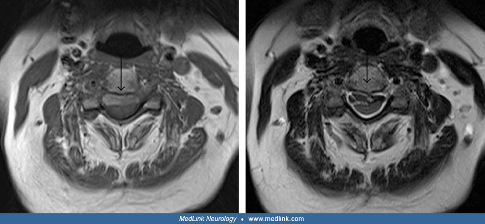
T2-weighted spine MRI in axial (a) and sagittal (b) section shows a longitudinally extensive epidural CSF collection from T2 to T8 (E) with a large spike-like osteophyte at T5/T6 impinging on the dura (G). The CT myelogram (c) shows contrast in the epidural collection from a CSF leak adjacent to the osteophyte (G). Legend: (A) spinal cord coated with hemosiderin; (B) epidural fat, bright on T2; (C) dura posterior to cord; (D) intradural CSF surrounding cord; (E) epidural CSF collection; (F) dura anterior to cord; (G) osteophyte; (H) CSF pulsation artifact. (Source: Halmagyi GM, Parker GD, Chen L, et al. Progressive loss of hearing and balance in superficial siderosis due to occult spinal dural defects. Eur Arch Otorhinolaryngol 2023;280[2]:633-41. Creative Commons Attribution 4.0 International [CC BY 4.0] license, creativecommons.org/licenses/by/4.0.)