Neuroimmunology
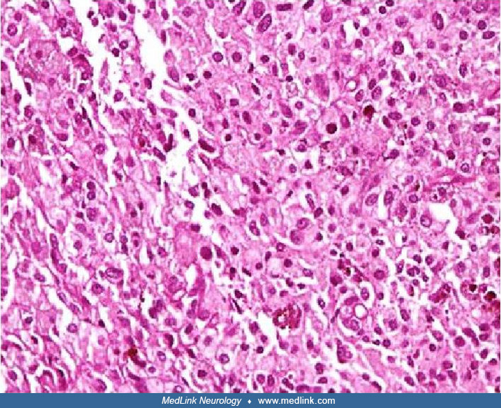
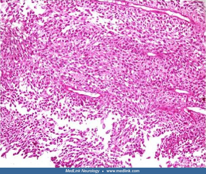
Congenital rubella
Dec. 13, 2025
MedLink, LLC
3525 Del Mar Heights Rd, Ste 304
San Diego, CA 92130-2122
Toll Free (U.S. + Canada): 800-452-2400
US Number: +1-619-640-4660
Support: service@medlink.com
Editor: editor@medlink.com
ISSN: 2831-9125
Toll Free (U.S. + Canada): 800-452-2400
US Number: +1-619-640-4660
Support: service@medlink.com
Editor: editor@medlink.com
ISSN: 2831-9125
Nearly 3,000 illustrations, including video clips of neurologic disorders.
Every article is reviewed by our esteemed Editorial Board for accuracy and currency.
Full spectrum of neurology in 1,200 comprehensive articles.
Listen to MedLink on the go with Audio versions of each article.
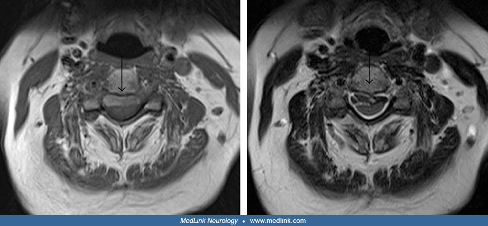
MRI in a 69-year-old woman with slowly progressive gait disturbances and hearing impairment. Sagittal T1-weighted MRI of the cervical and upper thoracic spine shows an anterior wedge degenerative deformation of the T6 vertebral body with a superimposed prominent central disc extrusion characterized by osteo-calcific signal (yellow arrow) causing a dural tear at the T6-T7 level. Above the disc extrusion, note the “sentinel” epidural fluid collection (red asterisk) stretching along the ventral aspect of the spinal canal and displacing the dura posteriorly. (Source: Bonomo G, Cusin A, Rubiu E, et al. Diagnostic approach, therapeutic strategies, and surgical indications in intradural thoracic disc herniation associated with CSF leak, intracranial hypotension, and CNS superficial siderosis. Neurol Sci 2022;43[7]:4167-73. Creative Commons Attribution 4.0 International [CC BY 4.0] license, creativecommons.org/licenses/by/4.0.)