General Neurology
Treatment of neurologic disorders with marijuana
Sep. 19, 2024
MedLink, LLC
3525 Del Mar Heights Rd, Ste 304
San Diego, CA 92130-2122
Toll Free (U.S. + Canada): 800-452-2400
US Number: +1-619-640-4660
Support: service@medlink.com
Editor: editor@medlink.com
ISSN: 2831-9125
Toll Free (U.S. + Canada): 800-452-2400
US Number: +1-619-640-4660
Support: service@medlink.com
Editor: editor@medlink.com
ISSN: 2831-9125
Nearly 3,000 illustrations, including video clips of neurologic disorders.
Every article is reviewed by our esteemed Editorial Board for accuracy and currency.
Full spectrum of neurology in 1,200 comprehensive articles.
Listen to MedLink on the go with Audio versions of each article.
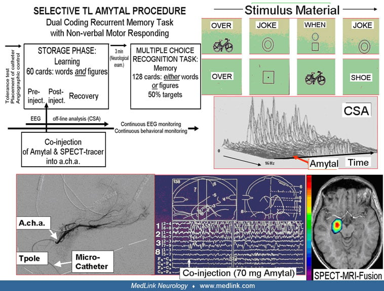
Illustration of the main components of the selective temporal lobe amobarbital memory test developed and used in Zurich. The graph (top, left) summarizes the test, which consists of a storage phase and a recognition phase (memory). The stimulus material is illustrated at the top, right. The storage phase consists of 60 cards, each containing both a word and a figure. In the recognition phase, 128 cards are presented that contain either a word or a figure. Half of them have been shown during the storage phase (= 50% targets). The patient responds nonverbally by button press (button ipsilateral to the injection). Via a transfemorally inserted microcatheter, the territory of the anterior choroidal artery is co-injected with amobarbital and a SPECT-tracer (bottom left, selective angiography of the AChA). During the STLAMT, the EEG is inspected on-line. The depicted example shows the appearance of slow delta activity in mesial temporal depth-EEG recordings (bottom middle). The EEG changes due to the amobarbital can be quantitatively analyzed and plotted to estimate the duration of the inactivation, for example, by compressed spectral array (CSA, middle portion, right margin). After termination of the amobarbital test, the SPECT is done. The SPECT image is then fused with MRI to exactly visualize the inactivated brain area (bottom, right). (Contributed by Dr. Heinz Gregor Wieser.)