Neuropharmacology & Neurotherapeutics
Methylphenidate
Jun. 17, 2021
MedLink, LLC
3525 Del Mar Heights Rd, Ste 304
San Diego, CA 92130-2122
Toll Free (U.S. + Canada): 800-452-2400
US Number: +1-619-640-4660
Support: service@medlink.com
Editor: editor@medlink.com
ISSN: 2831-9125
Toll Free (U.S. + Canada): 800-452-2400
US Number: +1-619-640-4660
Support: service@medlink.com
Editor: editor@medlink.com
ISSN: 2831-9125
Nearly 3,000 illustrations, including video clips of neurologic disorders.
Every article is reviewed by our esteemed Editorial Board for accuracy and currency.
Full spectrum of neurology in 1,200 comprehensive articles.
Listen to MedLink on the go with Audio versions of each article.
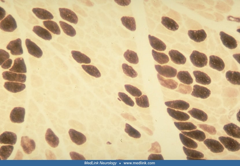
Longitudinal paraffin-embedded sections of vastus lateralis muscle of a child with nemaline myopathy. Hematoxylin-eosin (not shown) and Gomori trichrome (not shown) did not demonstrate nemaline rods, but they are clearly seen on formalin-fixed paraffin sections stained with phosphotungstic acid-hematoxylin (left), in which they appear deep purple (as do the Z-bands from which they are derived); and by the Luna-Parker acid fuscin stain (right), in which they appear bright pink against a pale green background of myofibrils. (x400) (Contributed by Dr. Susan Iannaccone.)