Neuroimmunology
Management of multiple sclerosis in COVID-19 pandemic
Feb. 13, 2026
MedLink, LLC
3525 Del Mar Heights Rd, Ste 304
San Diego, CA 92130-2122
Toll Free (U.S. + Canada): 800-452-2400
US Number: +1-619-640-4660
Support: service@medlink.com
Editor: editor@medlink.com
ISSN: 2831-9125
Toll Free (U.S. + Canada): 800-452-2400
US Number: +1-619-640-4660
Support: service@medlink.com
Editor: editor@medlink.com
ISSN: 2831-9125
Nearly 3,000 illustrations, including video clips of neurologic disorders.
Every article is reviewed by our esteemed Editorial Board for accuracy and currency.
Full spectrum of neurology in 1,200 comprehensive articles.
Listen to MedLink on the go with Audio versions of each article.
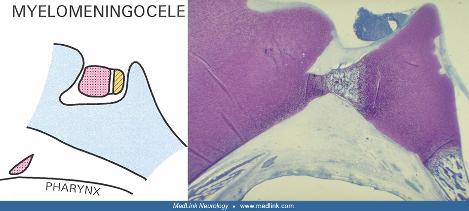
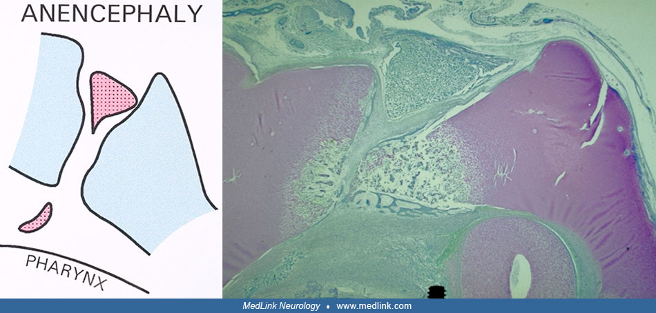
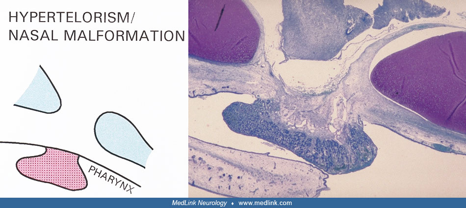
Left schematic drawing and right histological sagittal section of the sella turcica region and the pituitary gland from a normal human fetus, gestational age 16 weeks. Anterior direction is left. The adenopituitary gland appears dark blue (left) and the neuropituitary gland light blue (right). Ossification of the sella turcica appears in the cartilage (violet) below the pituitary gland. (Toluidine blue) (Reproduced with permission. J Craniofac Genet Dev Biol 1995;15:222-9.)