Neuroimmunology
Nonparaneoplastic autoimmune cerebellar ataxias
Mar. 28, 2026
MedLink, LLC
3525 Del Mar Heights Rd, Ste 304
San Diego, CA 92130-2122
Toll Free (U.S. + Canada): 800-452-2400
US Number: +1-619-640-4660
Support: service@medlink.com
Editor: editor@medlink.com
ISSN: 2831-9125
Toll Free (U.S. + Canada): 800-452-2400
US Number: +1-619-640-4660
Support: service@medlink.com
Editor: editor@medlink.com
ISSN: 2831-9125
Nearly 3,000 illustrations, including video clips of neurologic disorders.
Every article is reviewed by our esteemed Editorial Board for accuracy and currency.
Full spectrum of neurology in 1,200 comprehensive articles.
Listen to MedLink on the go with Audio versions of each article.
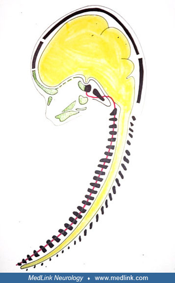
Histological sagittal section of a human embryo, gestational age 7 weeks. Anterior direction is left. The notochord appears in the cervical bodies and in a zigzag course in the cranial base, ending rostrally close to the developing pituitary gland. A pronounced metachromatic cartilaginous tissue is seen close to the notochord. (Toluidine blue) (Reproduced with permission. J Craniofac Genet Dev Biol 1995;15:157-61.)