


Sleep Disorders
Sudden infant death syndrome
Jul. 05, 2026
MedLink, LLC
3525 Del Mar Heights Rd, Ste 304
San Diego, CA 92130-2122
Toll Free (U.S. + Canada): 800-452-2400
US Number: +1-619-640-4660
Support: service@medlink.com
Editor: editor@medlink.com
ISSN: 2831-9125
Toll Free (U.S. + Canada): 800-452-2400
US Number: +1-619-640-4660
Support: service@medlink.com
Editor: editor@medlink.com
ISSN: 2831-9125
Worddefinition
At vero eos et accusamus et iusto odio dignissimos ducimus qui blanditiis praesentium voluptatum deleniti atque corrupti quos dolores et quas.
Tethered spinal cord is a condition in which spinal cord movement within the spinal column is limited. Excessive tension on the spinal cord leads to tethered cord syndrome, which can manifest as pain, motor or sensory deficits, and bowel and bladder dysfunction. Because most cases are primary or related to congenital spinal dysraphism, patients typically present in childhood, adolescence, or early adulthood (77; 54). Secondary, or acquired, etiologies of tethered cord syndrome include spinal cord neoplasm, infection, or fibrosis (34; 136); consequently, delayed presentation can occur (54). In October 2024, given the wide variation in current clinical practices, the Agency for Healthcare Research and Quality commissioned a systematic review of the diagnosis and treatment of tethered spinal cord, which provided evidence for the use of MRI or ultrasound for diagnosis (54). In symptomatic patients, surgical detethering improved neurologic outcomes and complications associated with open detethering surgery, while underscoring the need for stronger studies to guide diagnosis and treatment. Diagnosis of tethered cord syndrome is made primarily by clinical assessments and imaging. Although the need for surgical treatment is straightforward in some cases--for example, in a symptomatic patient with a low-lying conus and a fatty filum (131)--a case-by-case evaluation is necessary (77; 54). For instance, patients who undergo myelomeningocele closure appear tethered on MRI; however, only 40% require subsequent untethering (18). Furthermore, natural history and symptom progression rates differ among patients (54). In this article, the authors describe the etiology, pathogenesis, presentation, diagnosis, and treatment of tethered cord syndrome.
|
• Tethered cord syndrome describes a constellation of symptoms secondary to tethering of the spinal cord. | |
|
• Symptoms of tethered cord syndrome usually arise in childhood but sometimes present during adolescence or adulthood (104). | |
|
• When symptoms arise in adulthood, the underlying pathology is usually caused by occult spinal dysraphism (104). | |
|
• Occult tethered cord syndrome is increasingly being recognized, presenting with either pain or urological symptoms in adults or children, respectively. | |
|
• Failure to timely diagnose and treat tethered cord syndrome results in worse long-term outcomes. | |
|
• Neurologists are often the medical providers who make the diagnosis. | |
|
• Radiological diagnosis is confirmed with conus medullaris position below the L2-L3 disc space on MRI and may demonstrate filum terminale thickness greater than 2 mm. | |
|
• Early operative detethering is proposed by many neurosurgeons to prevent neurologic, orthopedic, or urological deficits from developing or worsening (104). | |
|
• A goal of initial surgery is to prevent recurrent tethering, which can occur in up to 50% of patients (104). |
In 1641, a Dutch physician named Nicholas Tulp was the first to use the term “spina bifida” (60). In 1761, Giovan Morgagni, the father of organ pathology, described a relationship between hydrocephalus and spina bifida (22). In 1857, A. Johnson described a young child with worsening lower extremity symptoms in whom a lesion consistent with a lipoma was found during exploratory surgery, and symptoms resolved when the spinal cord was freed from its dural attachments (61). In 1875, Virchow introduced the term “spina bifida occulta” (60).
The first untethering operation was reported by Jones in 1891 in a 22-year-old patient presenting with clubfoot deformities, lower extremity atrophy and pain, and voiding difficulties who underwent division of a “dense adventitious fibrous band” (63). The patient showed improvement 6 months after surgery. In 1910, Fuchs reported in patients with myelomeningocele incontinence induced by flexion (36).
In 1916, a neurologist, WG Spiller, first described the phenomenon of activity exacerbating symptoms in two adolescent patients after a rowing exercise, which highlighted the importance of early diagnosis in providing anticipatory guidance for activity avoidance (120).
In 1918, WM Brickner reported a correlation between early treatment and improved outcomes and proposed prophylactic treatment “in the hope of obviating the development of symptoms during adolescence” (20).
Different forms of spinal dysraphism were described over the next few decades, resulting in similar symptomatology (61; 20).
In 1940, Lichtenstein reported a relationship between spinal cord dysfunction and tethering lesions (78). In 1953, Garceau documented three patients with worsening spinal deformity and neurologic function, with reported recovery in patients with filum resection (38; 21; 31). Garceau hypothesized that a lesion in the filum terminale caused tension on the conus medullaris and coined filum terminale syndrome. Supporting evidence that the filum was under excessive tension came a few years later from Jones and Love, who reported recovery of neurologic and urologic functions after sectioning a tight filum and retracting the cut ends in six patients (62).
In 1976, Hoffman, Hendrick, and Humphreys published a landmark study and coined the term tethered spinal cord in patients with (1) a low-lying conus medullaris and (2) a thick filum terminale (diameter greater than 2 mm) (52). The study included 31 children, excluding those having obvious spinal dysraphisms, and documented variable presentations of tethered spinal cord, the role of myelography, making a diagnosis based on clinical suspicion despite normal imaging, and positive outcomes with surgical intervention. (52; 104). Since Hoffman and colleagues’ study, the definition of tethered spinal cord has been expanded to include various etiologies, and the pathophysiology has been proposed to involve mechanical traction, metabolic abnormalities, and changes in spinal cord blood flow (65). Studies rely mostly on animal models, which have limited ability to mimic human physiology (54).
In 1981, Yamada and colleagues showed that a tethered cord is linked with metabolic abnormalities by studying cytochrome c enzyme in mitochondria of humans and cats (145). They showed that increased traction on the spinal cord, induced by hanging weights, was associated with hypoxia, metabolic changes, and energy depletion; furthermore, the degree of metabolic impairment correlated with the severity of neurologic symptoms. Their results had strong implications for intervention: surgical untethering yielded a return to a normal metabolic state in mild or moderate redox changes and subsequent neurologic improvement. However, in more severe states, limited metabolic changes and neurologic changes were seen, suggesting that early intervention is key for prognosis (145; 143; 34). Research in the animal model was improved by Huang and colleagues, who produced chronic slow traction to more closely mimic the progressive nature of the disease (55). In 1987, Kang and colleagues demonstrated that untethering in kittens can improve spinal cord blood flow (65). However, in patients with tethered spinal cords compared with controls, no change in blood flow has been documented, and chronic ischemic injury has not been proven on a histological level (54).
Clinical evaluation and imaging remain the main criteria for diagnosis. Nonetheless, a normal appearance of the filum does not imply normal function. Selcuki and colleagues reported 13 patients with a normal level conus medullaris and incontinence who had improved continence after tight filum terminale resection (112; 113). As such, our current understanding of this syndrome is a complex lesion with heterogeneous causes (56; 02; 77; 142; 143). Operative care has advanced over the last few decades.
Tethered cord syndrome refers to a group of neurologic abnormalities involving the cauda equina and the spinal cord (54). Proposed pathophysiology involves multiple pathways that lead to chronic traction of the spinal cord, which can be in the setting of a collection of developmental anomalies of the caudal vertebrospinal axis or the consequence of their surgical repair (131; 54). As noted by Humphreys, the term is descriptive at best because its clinical presentations are as variable as its causes (56; 02; 77; 142). It does not denote (1) causation; (2) whether the dysraphism is open or closed; (3) at what level the cord is tethered; or (4) whether the causative agent is intradural, extradural, or both. There are two major groups of patients seen with this condition: (1) individuals with known spinal dysraphism who often have been previously operated on, and (2) individuals with a currently undiagnosed underlying spinal dysraphism. A neurologist is likely to encounter both types of patients. It is important to understand the clinical presentation of this condition, its proper evaluation, and when to consider referral to a neurosurgeon.
Tethered cord syndrome can present in childhood, and it most often does so in individuals with obvious spinal dysraphism, such as repaired myelomeningocele or lipo(myelo)meningocele. Clinical manifestations in children include progressive motor and sensory deterioration (particularly motor, with difficulties with gait or running); deterioration in urologic function; developmental or orthopedic deformities of the foot, leg, and spine; and, less commonly, back and leg pain (52; 72; 103; 45; 02; 21; 77; 142; 19; 51). In adolescents, scoliosis, pain, and sphincter dysfunction predominate. Symptoms may be exacerbated by back flexion and extension. There may be a history of prior orthopedic or urological surgeries, especially if the possibility of tethered cord syndrome was not appreciated (02; 21; 77; 19). Although long thought to be a disorder of children, adult presentations, and the usefulness of surgical detethering have become increasingly appreciated (74; 144; 75; 39; 40).
Pang and colleagues were the first group to present a series of adults with tethered cord syndrome along with clinicopathologic correlation (96). Over half of the patients in their series were without neurologic problems during childhood. The other 44% had only minor nondisabling problems in childhood, such as fixed-foot deformities or mild sensorimotor findings. They suggested that although the underlying pathologies between the childhood and adult syndromes were similar, the clinical manifestations and outcomes were different. Since then, numerous case series have shown that adults are much less likely to present with scoliosis or foot deformities. Pain and sensorimotor changes are much more common in adults than in children. The pain is most frequently back or leg pain, nondermatomal, shocklike or burning in quality, often with referral to the anorectal region, and can occur in up to 80% of patients. The straight-leg raising test may be positive in patients with tethered cord syndrome, which is consistent with the observation that symptoms may be exacerbated by back flexion and extension, and low back pain may be seen. Bladder or bowel symptoms are frequently seen, although less frequently than in most childhood series. Cutaneous stigmata of spinal dysraphism, such as lumbosacral subcutaneous lipoma, midline hypertrichosis, and sacral nevi, are relatively common (20% to 50% of patients) but are not universal. On neurologic exam, weakness may be subtle or pronounced, with involvement of multiple myotomes. Both upper and lower motor signs may be seen. With sensory exam, skip lesions are common. Reflexes and tone are variable and may have both upper and lower motor signs in different areas (96; 75; 02; 77; 39; 71; 40).
Tethered spinal cord can present as congenital scoliosis (99; 02; 77; 87; 107). This is important to identify because the tethered cord should be repaired prior to or simultaneously with scoliosis surgery (21; 19; 14). Although there are reports of children presenting with tethered cord as isolated urinary incontinence with features of neurogenic bladder (112), larger studies have failed to show increased risk for tethered cord in patients presenting only with lower urinary tract dysfunction (90), and other studies have shown the correlation of urinary incontinence to be greater with sacral dysplasia rather than tethered cord itself (01). Intractable constipation has also been reported as a predictor for tethered cord (105), but a report failed to show any increased risk for tethered cord in children with chronic constipation (17). Some children with urinary symptoms alone and normal neuroimaging may represent occult tethered cord, and these patients represent a therapeutic challenge because many of them will spontaneously improve without treatment, and surgery may result in clinical deterioration in some (127). Given the high frequency of urinary incontinence and constipation in children, and the fact that most patients will improve either spontaneously or with medical treatment alone, it is difficult to advocate surgery for patients with bowel or bladder symptoms and no other evidence of tethered cord (133; 28; 114; 19). Additionally, although terminal syringomyelia can frequently be associated with tethered cord, it appears to make no difference in outcome whether or not the syrinx is treated (16). Arachnoid cysts can also be found in association with spinal dysraphism but rarely require treatment (101).
Cutaneous stigmata are very common, seen in up to 60% of patients with tethered cord. These include hypertrichosis, cutaneous capillary hemangiomas, dermal sinus tracts, midline subcutaneous lipomas, lumbosacral skin appendages, and atretic meningocele “cigarette burn” lesions (02; 21; 77; 19; 25). However, a study showed that coccygeal pits are not associated with spinal dysraphism (137). It appears that a consensus has not yet been reached on what comprises the full spectrum of symptomatic tethered spinal cord. Even the definition of tethered cord is still being defined, given reports of a normal appearing filum terminale that can still exhibit histological abnormalities that lead to symptoms and respond to untethering (113; 114; 142). This is in part because accurate localization of the caudal tip of the spinal cord cannot be determined except surgically (neuroimaging is only an estimate), and filum thickness is less important than filum elasticity (142). Furthermore, some authors have speculated that some patients are genetically programmed to have a high conus and, thus, the position of the conus at a normal level is actually abnormal for that patient (122). Additionally, a normal filum but low conus medullaris (terminating below the L2-L3 disc space) should always be considered as tethered cord (67; 142).
Sometimes operative revision is needed after index tethered cord release. Any patient with a history of tethered cord syndrome or other spinal cord anomalies who has had prior surgery and who then presents with new symptoms may have retethering. This is unrelated to whether the original untethering procedure was for an asymptomatic or symptomatic condition (44). In fact, up to 25% of patients can experience retethering between 1 month and 20+ years after surgery (116). Surgery can arrest or even reverse these new symptoms, but careful monitoring is required to detect new symptoms (10; 06; 80; 116; 95).
A series study of 55 pediatric patients from a single medical center in Australia who underwent detethering surgery over a 5-year period reported that the most common complications were wound infection and cerebrospinal fluid leak, with six children requiring reoperation for wound issues and two patients for cord retethering during the study period. There were no deaths or new neurologic deficits. Of children with preoperative deficits, 26.7% were documented to have improvement or resolution of their symptoms postoperatively. The highest rate of improvement occurred in children with motor, gait, or sphincteric disturbances (126). Tethered cord release surgeries can also lead to the development of spinal intramural arachnoid cysts. Although uncommon, these cysts must be considered in the case of new compressive symptoms after an intradural procedure such as spinal cord untethering (42).
A review of surgical treatment of tethered cord syndrome in children in the United States demonstrated a steady trend of increasing operative treatment in more recent years. These studies demonstrated that older children tended to have more complications, longer lengths of stay, and higher hospital costs. Furthermore, the data supported surgical intervention at a younger age, in order to both improve outcome and reduce health care costs (118).
Data on adults suggest that surgery can be beneficial in improving symptoms, especially pain, but surgical intervention is advocated only in symptomatic patients (71; 40). Furthermore, surgical intervention in symptomatic adult patients has been reported to result in significant improvement and even reversal of neurologic deficits such as motor sensory dysfunction and bladder dysfunction (46; 37).
A 12-year-old girl presented to the spina bifida clinic. She previously underwent lumbar myelomeningocele repair in the neonatal period. She had a recent history of increased fatigue when walking more than a block. She also reported increased urinary leakage between her bladder catheterizations. There was no history of recent trauma.
Her exam showed some increased weakness of both ankles with dorsiflexors. Her patellar deep tendon reflexes were hyperactive, and her ankle reflexes were absent (as they often were in myelomeningocele). The tone in her lower extremities was moderately increased. There was a sensory deficit below L5. Her gait was somewhat Trendelenburg and crouched. She had a vertical surgical scar in the lumbosacral region of her back. MRI of her lower spine showed a tethered spinal cord with the conus medullaris ending at L5.
The patient underwent a spinal cord untethering. Postoperatively, she had improved endurance and ankle dorsiflexion strength. Since the untethering, her clinical improvement has remained stable, with no new symptoms or signs of a tethered spinal cord.
A basic understanding of the embryology of the central nervous system helps understand tethered cord syndrome. Tethered cord is thought to be a disorder of secondary neurulation with regulatory genes separate from those responsible for primary neurulation (15). Primary neurulation starts around gestational age 18 to 28 days, when the notochord induces the overlying ectoderm to form the neural plate, which later involutes to form neural folds that ultimately become a neural tube. At the same time, somites alongside the notochord form sclerotome cells, which later become the vertebral column.
The neural tube starts to close initially at the future cervical levels around 22 to 23 days of gestation and then at the caudal aspect around 26 to 27 days of gestation.
In a separate process called secondary neurulation, between 28 to 48 days of gestation, the distal remainder of the caudal tube forms via canalization, whereby undifferentiated cells from the primitive streak form a caudal cell mass that later forms vacuoles, fuses, and then forms a tube. Secondary neurulation forms the conus medullaris, cauda equina, and the filum terminale.
Around 43 to 48 days of gestation, the ventricular terminalis, the future conus medullaris, forms. The rostral and the caudal aspects later fuse (77; 48). During fetal life, the spinal cord extends to the sacral area at the coccygeal level. There is a progressive “ascent” of the conus medullaris relative to the vertebrae as the vertebral column grows out of proportion to the spinal cord (77). Defective retrogressive differentiation of the caudal neural tube may cause a tight filum (31). By 33 weeks of gestation, it is most commonly at or above the L2-L3 disc space, and by 2 months postnatally, it is at or above the L1-L2 disc space (13). In fact, MRI and ultrasound data suggest that the conus may be at the L1-L2 space as early as 40 weeks’ gestation (21). The shortening of the cord is secondary to retrogressive differentiation that results in the filum terminale, cauda equina, and ascension of the conus relative to the vertebral bodies; continued growth of the vertebral column causes further ascension of the conus, stretching of the filum, and growth of the cauda equina. A study showed that the conus medullaris most commonly terminates at the L1-L2 disc space and, in the absence of tethering, virtually never extends past the L2-L3 disc space (67).
Tethered cord may be congenital (primary) or acquired (secondary). Primary causes of tethered cord are associated with congenital anomalies (135; 132; 02; 21; 77; 89; 50), including myelomeningocele, split cord, caudal agenesis, meningocele manqué (spontaneously healed meningocele), fatty filum terminale, imperforate anus, dermal sinus, lipoma, pseudotails, and others. Furthermore, there are multiple developmental syndromes associated with tethered cord such as neurofibromatosis-1, Klippel-Feil syndrome, FG syndrome (short stature, large head, congenital hypotonia, developmental delay, and other anomalies), Klippel-Trenaunay-Weber syndrome, Dandy-Walker anomaly, the Ehlers-Danlos syndromes, Haberland syndrome, Chiari malformation (type 1 or 2), OEIS (omphalocele, exstrophy of the cloaca, imperforate anus, and spinal malformations with tethered cord), and VATER (vertebral malformations, imperforate anus, tracheoesophageal fistula, and renal-radial anomalies) or VACTERL syndrome (VATER with cardiac and limb defects). A report demonstrated tethered cord in 39% of children with VACTERL, 7.9% with anal atresia, and 2.4% with tracheoesophageal fistula (94). Secondary causes of tethered cord syndrome include infection, trauma, tumor, and fibrosis, which can disrupt the normal anatomy of the caudal spinal cord. In fact, anything that alters the normal relationship between the filum and conus may result in tethered cord (02; 77; 34). A study showed that adipose tissue on neuroimaging correlated with a low-lying conus and fatty filum, whereas nerve twigs on filum pathology correlated with abnormal urodynamics (125).
The connection between the filum and conus is important because it allows the cord to stretch during flexion and extension movements. However, although the filum stretches linearly with applied tension, the spinal cord elongates in a nonlinear fashion, and the caudal portion of the cord stretches the most when tension is applied (52). With increased linear stress, blood flow to the caudal cord is impaired, resulting in hypoxia and impaired oxidative metabolism (143; 51; 123). Because the degree of traction correlates with the amount of dysfunction, the damage goes from being initially reversible to irreversible with increased stretch or prolonged traction. This explains how adults can present with tethered cord syndrome: the tethering has always been present, but extrinsic factors outside the cord, such as increasing fibrosis of the filum; growth spurt during the late-teen and early-adult period; increased physical activity, especially with flexion and extension of the spine; or osteoarthritic spinal stenosis, which restricts movement of the cord and filum, can all result in increased stress to the caudal spinal cord (143; 51; 123). Patients with spinal dysraphism that limits cord movement are predisposed to developing tethered cord (111). Further, adults can present after a precipitating event (ie, disc herniation), suggesting that the adult tethered cord may be under just enough tension as to not cause symptoms, and any increased stretch or tension that reduces blood flow could lead to hypoxia and tethered cord syndrome (30). Even normal daily movements of the neck and back may cumulatively result in increased tension, causing symptoms to develop over the years. Also, lesions such as a dermal sinus, lipoma, tumor, scar tissue, or fibrous tissue can progress, resulting in later-onset symptoms (143; 51; 123).
Heterozygous missense mutations at VANGL1 and FUZZY have been indicated as risk factors for tethered spinal cord syndrome, as well as altered methylation of MGMT and maternal folic acid deficiency. Copy number variants have been found in the genomes of affected patients, which include the COX8C gene. COX8C is associated with metabolic disorders of the nervous system (148).
Tethered cord likely represents a multifactorial process due to a combination of genetic and environmental factors. As previously mentioned, tethered cord is associated with numerous congenital spinal cord and anorectal malformations and malformation syndromes, and several studies have shown linkage to various chromosomal regions. The true incidence and prevalence of occult spinal dysraphism are unknown because closed defects are often diagnosed with the onset of symptoms or incidentally (21; 116). Although the incidence of open neural tube defects has declined over the past three decades due to folic acid supplementation and termination of pregnancy with prenatal diagnosis (09; 24), the incidence of occult spinal dysraphism and tethered cord syndrome has increased due to wider availability of MR imaging (21; 51; 104; 121). The global prevalence of neural tube defects hit a plateau following mandatory fortification of enriched cereal grain products in the United States (138; 81) and is estimated to be two cases per 1000 births (64). Folic acid supplementation has had the greatest impact on reducing the incidence of myelomeningocele (117). However, due to inadequate folic acid supplementation, neural tube defects continue to occur (85). Because data suggest that open and closed neural tube defects may be generally related, siblings of patients with known neural tube defects are at higher risk (21). In Turkey, the prevalence of tethered cord syndrome was screened in primary school children with enuresis and estimated to be 0.1%, which likely underestimates true prevalence in the general population that also includes asymptomatic patients (11; 104).
Because all patients with myelomeningocele have tethered cord, folic acid supplementation could reduce the incidence of these patients. To reduce the risk of neural tube defects, the U.S. Centers for Disease Control and most countries recommend a healthy diet and folic acid supplementation of 400 µg per day periconceptionally (09; 43). Other neural tube defect risks include mothers who are very young or very old in age, toxic medications, obesity, and zinc deficiency (58). In the case of known disorders such as lipomas of the conus medullaris, preventative surgery before the first year of life is recommended to prevent future neurologic deterioration from subsequent spinal cord tethering (25). Furthermore, as discussed above, trauma, via either direct blows to the back or falls on the buttocks, to an already tethered cord can precipitate neurologic deterioration. Trauma should, therefore, be avoided, and the at-risk patient should be properly educated. Also, repeated or forceful stretching of an already stressed tethered cord could cause cord dysfunction. Such activities as straight-leg raising, forced forward bending, and prolonged sitting should be avoided or done carefully (141; 143; 51; 123).
With any case of neurologic deterioration in a patient with known spinal dysraphism, one must be suspicious of potential lesions throughout the entire neuroaxis and, thus, MRI of the entire spine should be performed (68). Deterioration in a patient who has previously undergone spinal surgery for detethering should be assumed to be due to tethered cord syndrome because scar tissue can cause (or worsen preexisting) tethered cord. Chiari malformation, syringohydromyelia, and shunt malfunction are possible reversible causes of symptoms that mimic tethered cord syndrome (21). In older age groups, both lumbar spinal stenosis and lumbosacral disc disease can cause symptoms suggesting tethered cord syndrome (26). Patients who do not meet imaging criteria for tethered cord syndrome (conus lower than the L2-3 disc space) may still have tethered cord due to other lesions (eg, filum lipoma) and may benefit from surgery (134). However, patients with normal imaging, referred to as occult tethered cord syndrome, represent a controversial population; certainly other causes for symptoms should be explored before performing detethering on these patients (21; 29; 114; 122; 127).
Case reports of patients presenting with tethered cord syndrome resembling plantar fasciitis and peripheral neuropathy (08), resembling Charcot-Marie-Tooth disease (119), or due to arteriovenous fistula (110; 83), have been reported in adults. Furthermore, pseudo tethered cord has been reported after lumbar puncture (93).
Cases with known dysraphic states. Any neurologic deterioration in a person with any type of known dysraphic state should be considered secondary to an underlying reversible condition. Each case should be aggressively evaluated with an eye towards identifying any potentially surgically correctable lesions such as tethered cord, syringohydromyelia, or hydrocephalus. It is not always clear whether once a lesion is found that surgical correction will reverse the symptoms. Implied in this approach is the need for a detailed assessment of the individual's baseline neurologic status and anatomy on which future potential changes can be looked for. Unfortunately, this is not always possible, and a "clinically educated" decision is the best that can be offered. A history of recent, rather than past or longstanding neurologic deficits, increases the likelihood of reversibility (21; 47; 19; 39). This is consistent with the model that reversible symptoms progress to irreversible with prolonged time between symptom onset and surgical correction. However, even patients with multiple prior surgical revisions or longstanding symptoms may still show neurologic improvement or reduction in pain with surgery (06; 46).
The conditions that are frequently associated with neurologic deterioration caused by cord tethering are myelomeningoceles, lipomyelomeningoceles, spinal or filum lipomas, split cord malformations, short, thickened filum terminale (greater than 2 mm in diameter), and fibrous bands that sometimes continue through the dura to bone and result in the dimpling of overlying skin. Other conditions include adhesions of the filum, conus, and nerve roots to the posterior or lateral dura; congenital dermoid cysts and sinuses; neuroenteric cysts; and sacral dysplasia, which can result in cord tethering, sometimes additionally with lumbosacral stenosis. Furthermore, if the patient undergoes spinal surgery for repair of a myelomeningocele or other tethering lesion, for example, later neurologic deterioration may be due to scarring from the surgery, resulting in adhesion and tethering of the cord (21; 19). All patients with myelomeningocele have a tethered cord at birth and are detethered at the time of myelomeningocele closure, typically within the first 72 hours of life. Subsequent detethering surgery for presumed retethering is done only if patients develop symptoms of tethered cord syndrome (106; 109); therefore, these patients require frequent follow-up, usually with a multidisciplinary clinic.
Baseline neurologic status is assessed with a complete neurologic history and physical exam. Particular attention should be paid to thorough strength (manual motor testing) and sensory examination and assessment of perineal function (bladder and bowel function, including cremasteric reflex and anal wink). It is often helpful to assess urologic status with urodynamic studies. Many authors recommend preoperative and postoperative urodynamic testing because it offers objective evidence of improvement after surgery and also provides a means for early detection of recurrent symptoms (149; 02; 77; 90; 35; 146). Even patients with very mild symptoms can have abnormal urodynamics (53; 80; 35). Detrusor overactivity is considered the most common finding when evaluating urologic status in tethered cord syndrome (129). This is particularly important because tethered cord syndrome remains a clinical and anatomic diagnosis. A 2018 Delphi study developed consensus statements among a group of experts regarding the clinical management of children with tethered spinal cords (05).
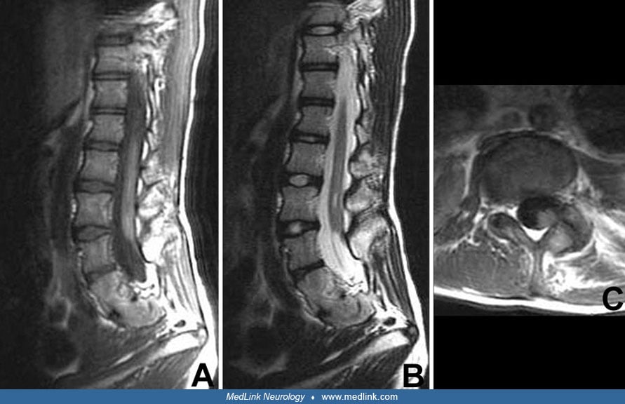
With the widespread availability of MRI, the evaluation of lumbosacral anatomy has become noninvasive and detailed (87; 107). MRI easily demonstrates the filum thickness and whether or not the conus ends at the proper place, or if it is caudally displaced. Placement of the conus below the L2-L3 interspace is consistent with tethered cord, with filum thickness often greater than 2 mm (02; 77). A small percentage of patients with tethered spinal cord have been demonstrated to have a normally positioned conus, called an occult tethered cord; however, these patients always have clincal symptoms consistent with a tethered spinal cord, and they often have other associated radiographic abnormalities, such as filum lipomas, syrinx, or congenital vertebral body anomalies that are identifiable on MRI (134; 128; 114). As the quality of MRI improves, the sensitivity for identifying filum abnormalities will continue to improve (33).
We are left with the following conclusion: MRI imaging can tell us in the vast majority of circumstances whether a spinal cord is anatomically tethered, but clinical history, symptoms, and examination are critical in making surgical decisions regarding intervention for a tethered cord.
Cases that present without a known underlying dysraphic state. Unlike the patient with a known dysraphic state, who is likely to present to a multidisciplinary spina bifida clinic, some patients present with symptomatic tethered cord with no known underlying (or occult) dysraphic state. These patients can present in many settings, not least of which could be a neurologist's office. Usually, these patients develop symptoms during childhood, but they can become symptomatic at an earlier age, during adolescence, or in adult life. Neurologic dysfunction is often slowly progressive. Symptoms and signs can appear stable over serial exams, sometimes for years. However, even within this seemingly stable setting, minor neurologic deficits may progress significantly and suddenly following minor traumatic events (96; 46). Neurologic dysfunction manifests itself by lower extremity paresis and deformity, spinal deformity, sensory loss (including trophic ulceration), and changes in bladder and bowel function (32; 45; 75; 02; 77). Dysfunctional bladder may be the only presenting manifestation in adults (72), although this is not universally accepted (133; 29; 114). Although urinary incontinence in children has not been shown to be predictive of occult spinal dysraphism (90), children with occult tethered cord tend to present more often with urinary symptoms, in contrast to adults who present more often with pain complaints (127). Orthopedic deformities include progressive scoliosis and pes cavus, varus, or valgus deformities of the foot, or leg-length discrepancies (02; 21; 77; 51; 41). Most often, these deformities are unilateral, and progressive atrophy of the lower extremities can be seen. Also, patients with anorectal and urogenital malformations have a higher incidence of tethered cord syndrome (135; 21; 51; 94). Unless a clear spinal dysraphism is identified, however, these children should be treated medically rather than surgically (27).
Workup of these individuals may include the following (02; 21; 77; 51):
• Lumbosacral plain x-rays. Some cases of tethered cord syndrome have lumbosacral spina bifida that can be detected on plain radiographs. | |
• Anorectal malformations can include dermal sinus tract, gluteal cleft deviation. Split cord malformations can cause tethered cord syndrome; for example, type I split cord malformation demonstrates a bony spicule dividing the two hemicords. | |
• MRI of the lumbosacral spine with sagittal and axial views. This is by far the most sensitive and specific way of identifying cases of tethered cord syndrome. Cord termination below L2-3 disc space is consistent with tethered cord (147; 02; 21; 67; 77; 51). A normally ending conus at L1-L2 may still be of concern if the cord appears taut or if there are other associated abnormalities (lipoma, fatty filum) and the patient is symptomatic (134; 128; 114; 122; 142). Use of the prone position during MRI may increase the sensitivity for detecting occult tethered cord (88), but this is controversial. | |
• CT with or without myelography may be useful, particularly for demonstrating bony abnormalities and determining the nature of the septum in split cord malformations; however, myelography is seldom needed or used due to being invasive and difficult to perform in infants, and also because of the associated radiation dose (21). | |
• Ultrasound is useful during the prenatal and newborn period for demonstrating the level of the conus. It can serve as a screening tool up to about 2 months of age and confirmed by MRI (84; 21; 49). There has been literature suggesting the overuse of ultrasounds in asymptomatic patients presenting with simple sacral dimples to assess the possibility of tethered spinal cord. Sacral dimples are common and the presence of abnormalities with sacral dimples was equal to those without. Ultrasounds for these patients is not warranted (03). | |
• Urodynamics should be performed to assess bladder function before and after surgery and also may be helpful in cases where neuroimaging is normal (75; 21; 35; 146). | |
• Assessment of anorectal function may be useful, but is not widely measured (66). | |
• Width of the P37 peak of the posterior tibial nerve somatosensory evoked potential has been reported as a useful marker for tethered cord syndrome (76), but this type of testing is impractical and infrequent. Those with a normal somatosensory evoked potential may not require further investigation with MRI (124). |
State-of-the-art management of tethered cord syndrome includes early identification (particularly in high-risk individuals with spinal dysraphism), appropriate decisions regarding indications for surgical intervention, and close postoperative follow-up. Although the decision to operate is easy in symptomatic patients with spinal dysraphism, it is not always clear when to operate in asymptomatic patients. In a review, Yaltirik and colleagues proposed that asymptomatic patients with non-spina bifida associated tethered cord syndrome and concomitant scoliosis may benefit from fusion with osteotomies without untethering and should be followed conservatively as scoliosis patients (140).
In terms of the surgical procedure, primary attention is paid to releasing the stretched spinal cord. However, clinicopathologic correlations have shown that not all of the actual etiology for tethering can be noted preoperatively (102; 142; 139). The point to emphasize is the importance of having a neurosurgeon with operative experience with tethered cord syndrome involved in managing the case. Most authors or neurosurgeons now believe that infants with lipomeningocele and a tethered cord should be operated on in the first few months of life before they become symptomatic. Certainly, symptomatic children should be operated on, preferably when symptoms are absent or mild (02; 21; 77; 19; 126). Although surgery in adults may involve a greater risk of neurologic injury than in children (especially in the elderly), and benefit may be reduced, especially if the symptoms have been longstanding, it is generally a low-risk procedure with significant benefit in carefully selected patients (39; 92). Because neurologic deficits from a tethered spinal cord may be irreversible, early surgery is recommended (59; 57; 75; 47; 39). Minimally invasive approaches have been used, and outcome data appear similar to the traditional open approach (97). Spinal column shortening has been proposed as a safer alternative to direct spinal cord untethering in complex tethered spinal cords, with favorable outcomes and fewer complications, but further research is required to establish efficacy (04). Intraoperative monitoring is also frequently used to help reduce the incidence of neurologic injury and has essentially become the standard of care (02; 77; 98; 108).
For postoperative care, a short period of flat-bed rest post-untethering is recommended. Prophylactic acetazolamide is occasionally administered to reduce CSF production and prevent leakage in untethering surgeries, but has been shown to be unnecessary and possibly harmful due to the side effects (115).
Certainly, improvement in urodynamics is important in deciding which patients should undergo surgery, and there have been efforts in predicting which patients are more likely to improve (35), as well as attempts to understand the threshold for surgery in patients with occult tethered cord and urinary symptoms (146; 07). This is especially important, given reports of spontaneous improvement in urodynamics in children with congenital spinal lipomas, a known cause of tethered cord (12). This also correlated with data that filum terminale lipomas may actually spontaneously disappear, and if found in asymptomatic patients, should be considered incidental findings according to the authors (23).
For those patients with recurrent tethered cord, pain management is of concern when further untetherings pose unlikely improvement and increased risk. Neuromodulation has been suggested as an alternative therapeutic option to achieve pain relief. Implantation of a dorsal spinal cord stimulator can give relief from neuropathic pain secondary to recurrent tethered spinal cord (130; 91).
Neurologic outcomes after cord untethering are generally good. Adults may not improve as much as children (96), although the more important factor may be the length of symptoms. Surgery during the asymptomatic period or when symptoms are mild may result in better outcomes (75; 02; 47; 77; 19; 51). However, even late release can result in improvement for some patients (46). In adults, pain symptoms improve the most following surgery, although the majority of patients also have improvement or stabilization in motor strength and bladder function (96; 57; 75; 39). In children, pain is a less common symptom, but most patients have stabilization or improvement in strength and bladder function (02; 21; 77; 19; 82). Grading systems to assess the degree of intraoperative untethering have been published (70), along with scales to rate clinical symptoms pre-and post-untethering (58) and attempts to better classify various subtypes of tethering (102). Outcome depends on the pathology and severity of symptoms. A retrospective study has shown that those with diastematomyelia and thickened filum presenting with tethered spinal cord have a better outcome than those with myelomeningocele or meningocele (58). Several studies have addressed the need for operative revision after index tethered-cord release if signs and symptoms of retethering develop (10; 133; 75; 06; 80; 116; 95).
A study demonstrated features on MRI following tethered cord release that may be useful in monitoring for change and detecting early recurrence of retethering (69). Another study reported improvement in intraoperative transcranial electrical motor-evoked potentials, although there was no correlation with long-term outcomes (100).
An overview of tethered cord surgeries in the United States demonstrated mortality rate, complication rate, and length of stay as 0.0005%, 9.48%, and 5.6 days, respectively (73). Complications from surgery can include hemorrhage/hematoma; infections, including meningitis; deep venous thrombosis or pulmonary embolism; neurologic decline/injury; retethering; and CSF leak (02; 77).
The most common complication of tethered cord syndrome in pregnancy is urinary tract infection. In 10% of pregnancies, hydronephrosis, intestinal obstruction, and renal complications occur due to the presence of the fetus. Caesarean section is often considered due to pelvic bony abnormalities, but vaginal birth may be performed as well. There is an increased risk for newborns to have neural tube deficits (4% to 7%), and 4.0 mg of folate is recommended every day to prevent neural tube deficits (86).
Symptoms of tethered cord syndrome can become worse after childbirth in the lithotomy position (96). Therefore, care needs to be taken with a patient with known spinal dysraphism.
Unless surgery is indicated shortly after birth, MRI is often delayed until at least 3 months of age so that sedation may be utilized for optimal imaging in infants when evaluating patients with occult spinal dysraphism (129).
In patients with a history of tethered spinal cord, the conus medullaris ends at a lower level, even extending down to L5-S1. Spinal anesthesia administered through a dural puncture should be avoided due to the possibility of serious injury to the spinal cord. Secondary hematoma can lead to a compression injury and possibly irreversible damage (79).
All contributors' financial relationships have been reviewed and mitigated to ensure that this and every other article is free from commercial bias.
Richard Anderson MD
Dr. Anderson of NYU Grossman School of Medicine has no relevant financial relationships to disclose.
See Profile
Bernard L Maria MD
Dr. Maria of Thomas Jefferson University has no relevant financial relationships to disclose.
See Profile
Bernard L Maria MD
Dr. Maria of Thomas Jefferson University has no relevant financial relationships to disclose.
See ProfileNearly 3,000 illustrations, including video clips of neurologic disorders.
Every article is reviewed by our esteemed Editorial Board for accuracy and currency.
Full spectrum of neurology in 1,200 comprehensive articles.
Listen to MedLink on the go with Audio versions of each article.
MedLink, LLC
3525 Del Mar Heights Rd, Ste 304
San Diego, CA 92130-2122
Toll Free (U.S. + Canada): 800-452-2400
US Number: +1-619-640-4660
Support: service@medlink.com
Editor: editor@medlink.com
ISSN: 2831-9125
Sleep Disorders
Jul. 05, 2026
General Child Neurology
Jun. 24, 2026
General Child Neurology
Jun. 10, 2026
Epilepsy & Seizures
Jun. 02, 2026
General Neurology
May. 13, 2026
General Child Neurology
May. 12, 2026
Developmental Malformations
May. 08, 2026
Developmental Malformations
May. 08, 2026