Headache & Pain
Migraine: pathogenesis and pathophysiology
Aug. 24, 2024
MedLink®, LLC
3525 Del Mar Heights Rd, Ste 304
San Diego, CA 92130-2122
Toll Free (U.S. + Canada): 800-452-2400
US Number: +1-619-640-4660
Support: service@medlink.com
Editor: editor@medlink.com
ISSN: 2831-9125
Toll Free (U.S. + Canada): 800-452-2400
US Number: +1-619-640-4660
Support: service@medlink.com
Editor: editor@medlink.com
ISSN: 2831-9125
Nearly 3,000 illustrations, including video clips of neurologic disorders.
Every article is reviewed by our esteemed Editorial Board for accuracy and currency.
Full spectrum of neurology in 1,200 comprehensive articles.
Listen to MedLink on the go with Audio versions of each article.
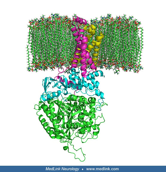
Ribbon diagram (Richardson diagram) of complex II. The structure of the complex II (succinate dehydrogenase or succinate-ubiquinone oxidoreductase) bound to the inner phospholipid membrane of mitochondria. Complex II is a heterotetramer with four dissimilar subunits, two of which are hydrophilic (SdhA and SdhB), whereas the other two are hydrophobic membrane anchor subunits (SdhC and SdhD). The mitochondrial inter-membrane space is at the top of the image. Legend: SdhA is green, SdhB is teal, SdhC is fuchsia, and SdhD is yellowish green. Phospholipids surround the membrane subunits. Illustration by Richard Wheeler (Zephyris), 2006. (Courtesy of Wikimedia Commons. GNU Free Documentation License.)