Behavioral & Cognitive Disorders
Alzheimer disease
Nov. 14, 2023
MedLink®, LLC
3525 Del Mar Heights Rd, Ste 304
San Diego, CA 92130-2122
Toll Free (U.S. + Canada): 800-452-2400
US Number: +1-619-640-4660
Support: service@medlink.com
Editor: editor@medlink.com
ISSN: 2831-9125
Toll Free (U.S. + Canada): 800-452-2400
US Number: +1-619-640-4660
Support: service@medlink.com
Editor: editor@medlink.com
ISSN: 2831-9125
Nearly 3,000 illustrations, including video clips of neurologic disorders.
Every article is reviewed by our esteemed Editorial Board for accuracy and currency.
Full spectrum of neurology in 1,200 comprehensive articles.
Listen to MedLink on the go with Audio versions of each article.
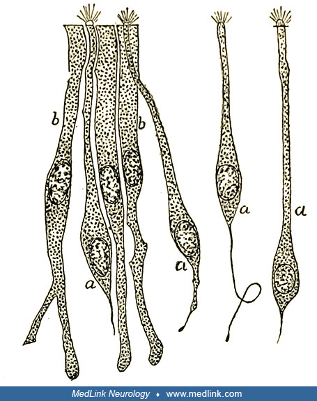
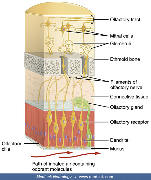
Santiago Ramón y Cajal's drawing of an anteroposterior section of the olfactory bulb of the duck. Legend: (A) arborization of olfactory fibrils within the glomeruli. (B) Descending trunks of the large tufted cells, terminating in a granulous meshwork in the glomeruli. (C) Large superior tufted cells. (Source: Ramón y Cajal S. Origen y terminación de las fibras nerviosas olfatorias [Origin and termination of the olfactory nerve fibers]. Gaceta Sanitaria de Barcelona 1890-1891:3; 133-139, 174-181, 206-212. See: Shepherd GM, Greer CA, Mazzarello P, Sassoè-Pognetto M. The first images of nerve cells: Golgi on the olfactory bulb 1875. Brain Res Rev. 2011;66[1-2]:92-105. Public domain.)