Peripheral Neuropathies
Diabetic neuropathies
Apr. 09, 2023
MedLink®, LLC
3525 Del Mar Heights Rd, Ste 304
San Diego, CA 92130-2122
Toll Free (U.S. + Canada): 800-452-2400
US Number: +1-619-640-4660
Support: service@medlink.com
Editor: editor@medlink.com
ISSN: 2831-9125
Toll Free (U.S. + Canada): 800-452-2400
US Number: +1-619-640-4660
Support: service@medlink.com
Editor: editor@medlink.com
ISSN: 2831-9125
Nearly 3,000 illustrations, including video clips of neurologic disorders.
Every article is reviewed by our esteemed Editorial Board for accuracy and currency.
Full spectrum of neurology in 1,200 comprehensive articles.
Listen to MedLink on the go with Audio versions of each article.
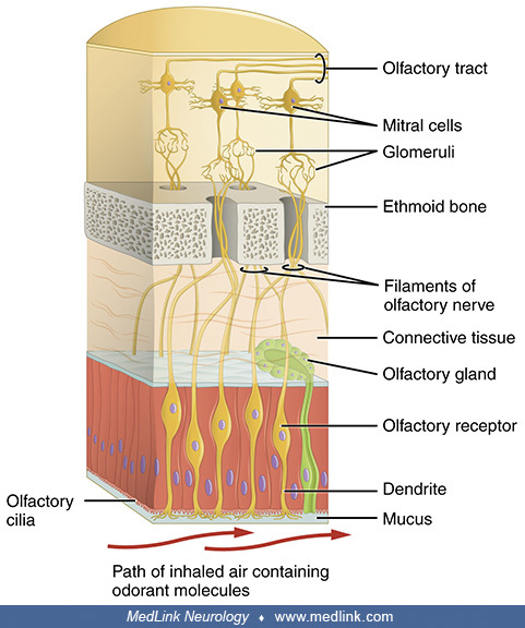
Immunohistochemical staining for Dab1 protein (reelin signaling mediator, green) and Map2a,b protein (microtubule-associated protein, red) on mouse olfactory bulb sections at P3. Nuclei counterstained with Hoechst (blue). Legend: (D) Olfactory nerve layer. (E) Olfactory glomerular layer. (F) External plexiform layer. (G) Mitral cell body layer. (H) Internal plexiform layer. (I) Granule layer. (Source: Martín-López E, Blanchart A, De Carlos JA, López-Mascaraque L. Dab1 (Disable homolog-1) reelin adaptor protein is overexpressed in the olfactory bulb at early postnatal stages. PLoS One 2011;6[10]:e26673. See also: Figueres-Oñate M, Gutiérrez Y, López-Mascaraque L. Unraveling Cajal's view of the olfactory system. Front Neuroanat 2014;8:55. Creative Commons Attribution [CC BY] license, creativecommons.org/licenses/by/4.0.)