Sleep Disorders
Non-24-hour sleep-wake disorder
Mar. 01, 2024
MedLink®, LLC
3525 Del Mar Heights Rd, Ste 304
San Diego, CA 92130-2122
Toll Free (U.S. + Canada): 800-452-2400
US Number: +1-619-640-4660
Support: service@medlink.com
Editor: editor@medlink.com
ISSN: 2831-9125
Toll Free (U.S. + Canada): 800-452-2400
US Number: +1-619-640-4660
Support: service@medlink.com
Editor: editor@medlink.com
ISSN: 2831-9125
Nearly 3,000 illustrations, including video clips of neurologic disorders.
Every article is reviewed by our esteemed Editorial Board for accuracy and currency.
Full spectrum of neurology in 1,200 comprehensive articles.
Listen to MedLink on the go with Audio versions of each article.
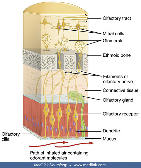
Cajal's drawing of the frontal section of the rabbit olfactory bulb, showing the layered distribution of cells. Legend: (1) Olfactory nerve layer. (2) Glomerular layer. (3) External plexiform layer. (4) Mitral cells layer. (5) Inner plexiform layer. (6) Granular and white matter layer. (a) Peripheral tufted cells, (b) middle; (c) internal (d) short axon cells. (Source: Ramón y Cajal S. Estudios sobre la corteza cerebral humana. IV. Estructura de la corteza cerebral olfativa del hombre y mamíferos. Trab Lab Invest Biol 1901;1:1-140. Instituto Cajal, Madrid, Spain. Figueres-Oñate M, Gutiérrez Y, López-Mascaraque L. Unraveling Cajal's view of the olfactory system. Front Neuroanat 2014;8:55. Creative Commons Attribution [CC BY] license, creativecommons.org/licenses/by/4.0.)