Sleep Disorders
Sleep disorders
Apr. 20, 2025
MedLink, LLC
3525 Del Mar Heights Rd, Ste 304
San Diego, CA 92130-2122
Toll Free (U.S. + Canada): 800-452-2400
US Number: +1-619-640-4660
Support: service@medlink.com
Editor: editor@medlink.com
ISSN: 2831-9125
Toll Free (U.S. + Canada): 800-452-2400
US Number: +1-619-640-4660
Support: service@medlink.com
Editor: editor@medlink.com
ISSN: 2831-9125
Nearly 3,000 illustrations, including video clips of neurologic disorders.
Every article is reviewed by our esteemed Editorial Board for accuracy and currency.
Full spectrum of neurology in 1,200 comprehensive articles.
Listen to MedLink on the go with Audio versions of each article.
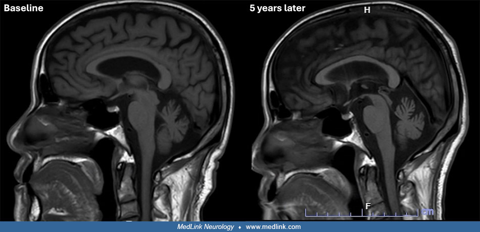
Midsagittal brain MRI of a patient with spinocerebellar ataxia type 27B reveals pure cerebellar atrophy, most pronounced at the vermis, at baseline (left) and with little interval change after 5 years (right). This illustrates the relatively slow progression of spinocerebellar ataxia type 27B compared with other repeat-expansion spinocerebellar ataxia. (Contributed by Dr. Pichet Termsarasab.)