Neuropharmacology & Neurotherapeutics
Phenytoin
Sep. 13, 2021
MedLink, LLC
3525 Del Mar Heights Rd, Ste 304
San Diego, CA 92130-2122
Toll Free (U.S. + Canada): 800-452-2400
US Number: +1-619-640-4660
Support: service@medlink.com
Editor: editor@medlink.com
ISSN: 2831-9125
Toll Free (U.S. + Canada): 800-452-2400
US Number: +1-619-640-4660
Support: service@medlink.com
Editor: editor@medlink.com
ISSN: 2831-9125
Nearly 3,000 illustrations, including video clips of neurologic disorders.
Every article is reviewed by our esteemed Editorial Board for accuracy and currency.
Full spectrum of neurology in 1,200 comprehensive articles.
Listen to MedLink on the go with Audio versions of each article.
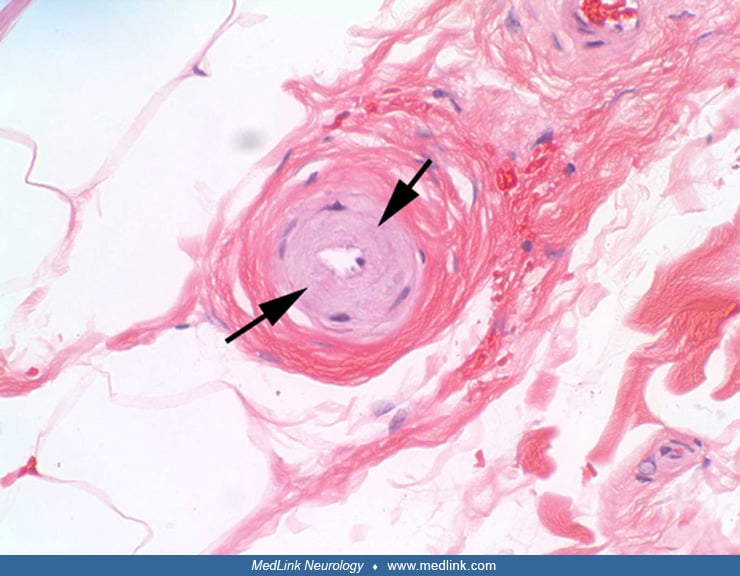
Electron microscopy reveals the deposition of granular osmiophilic material (arrows) and degeneration of a smooth muscle cell (Sm) in the media of the vessel wall. An endothelial cell (E) is lining the vessel lumen (L). (Contributed by Dr. Steve Moore and Dr. Jill Hagenkord of the University of Iowa Department of Pathology.)