Alpha-ketoglutarate dehydrogenase deficiency
Dec. 01, 2025
MedLink, LLC
3525 Del Mar Heights Rd, Ste 304
San Diego, CA 92130-2122
Toll Free (U.S. + Canada): 800-452-2400
US Number: +1-619-640-4660
Support: service@medlink.com
Editor: editor@medlink.com
ISSN: 2831-9125
Toll Free (U.S. + Canada): 800-452-2400
US Number: +1-619-640-4660
Support: service@medlink.com
Editor: editor@medlink.com
ISSN: 2831-9125
Nearly 3,000 illustrations, including video clips of neurologic disorders.
Every article is reviewed by our esteemed Editorial Board for accuracy and currency.
Full spectrum of neurology in 1,200 comprehensive articles.
Listen to MedLink on the go with Audio versions of each article.
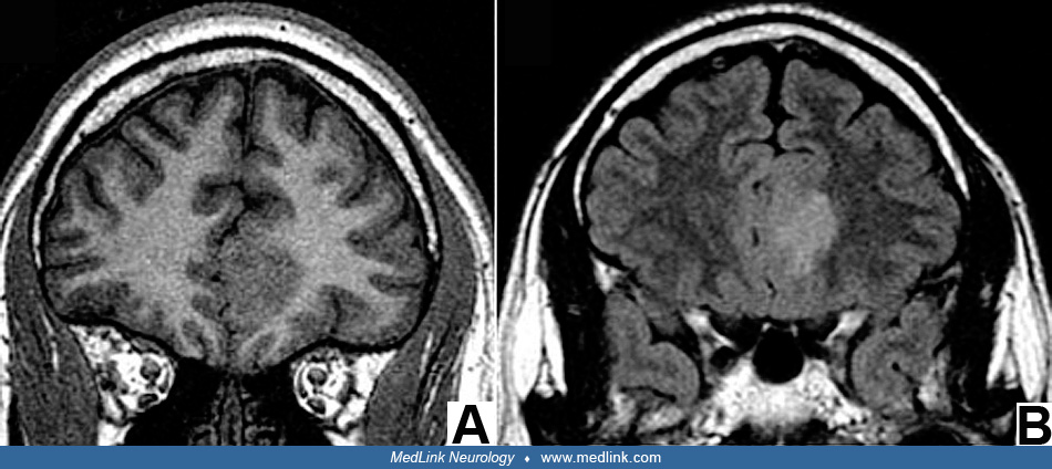
Coronal T2 (A), spoiled gradient recall (B), and FLAIR (C) weighted sequences demonstrate a focal area of cortical thickening (arrow) and abnormal signal in the left fusiform gyrus of the temporo-occipital lobe in a patient with refractory seizures. This area was resected and found to be consistent with focal cortical dysplasia pathologically. (Contributed by Dr. Diana Gomez-Hassan.)