Neuro-Ophthalmology & Neuro-Otology
Otic capsule dysplasia
Dec. 01, 2025
MedLink, LLC
3525 Del Mar Heights Rd, Ste 304
San Diego, CA 92130-2122
Toll Free (U.S. + Canada): 800-452-2400
US Number: +1-619-640-4660
Support: service@medlink.com
Editor: editor@medlink.com
ISSN: 2831-9125
Toll Free (U.S. + Canada): 800-452-2400
US Number: +1-619-640-4660
Support: service@medlink.com
Editor: editor@medlink.com
ISSN: 2831-9125
Nearly 3,000 illustrations, including video clips of neurologic disorders.
Every article is reviewed by our esteemed Editorial Board for accuracy and currency.
Full spectrum of neurology in 1,200 comprehensive articles.
Listen to MedLink on the go with Audio versions of each article.
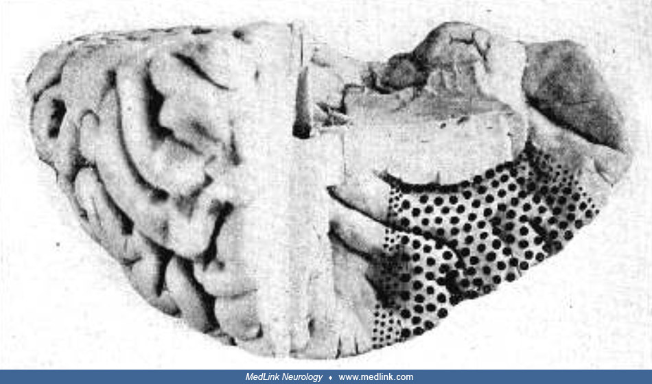
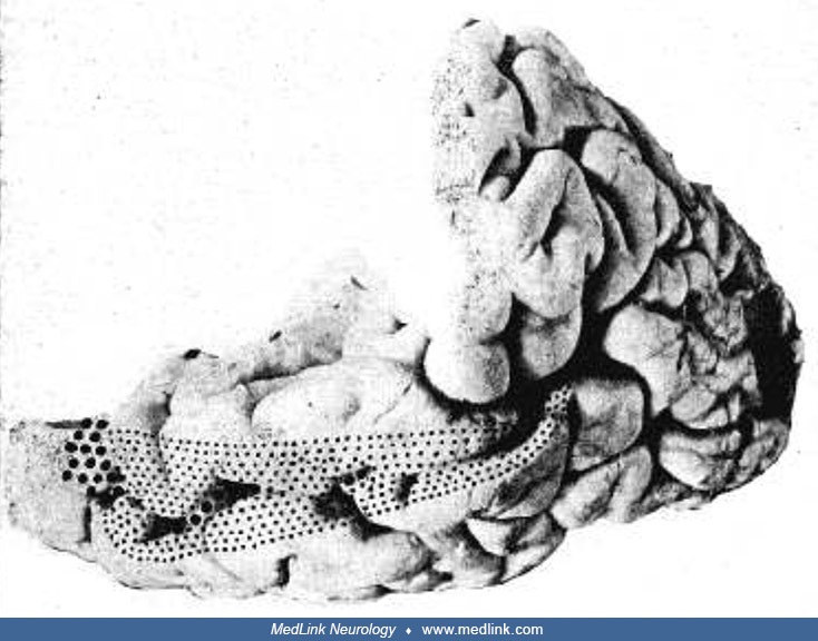
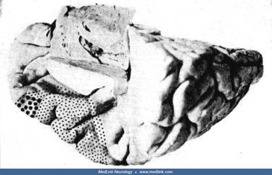
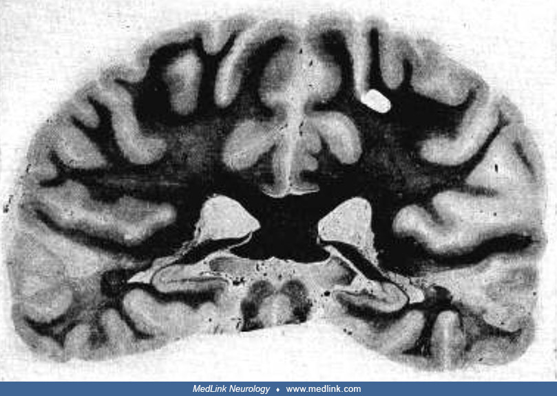
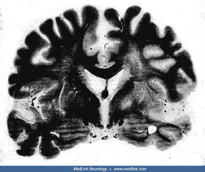
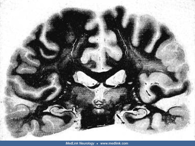
Coronal section of the brain, passing through the extreme posterior end of the transverse convolutions and insula, and through the geniculate bodies. (Left side) On the left side, the cortex of the ventral surface of the superior temporal gyrus is defective. The cortex of the upper surface of the middle temporal gyrus and most of the fibers of the center of the convolution are absent. The deep fiber areas of the lobe show beneath the bottom of T, a great diminution in numbers, and the degeneration continues inward as a thin streak traceable as far as the lenticular nucleus. This streak occupies the position of the fiber radiations between the medial geniculate body and the first temporal convolution. (Right side) The condition on the right side is much the same. The medial geniculate bodies on both sides give no evidence of degeneration changes; their cells are of normal appearance, and their fibers are abundant and deeply stained. (From: Barrett A. A case of pure word-deafness with autopsy. J Nerv Ment Dis 1910;37[2]:84.)