Peripheral Neuropathies
Hereditary amyloid polyneuropathy
Jan. 20, 2026
MedLink, LLC
3525 Del Mar Heights Rd, Ste 304
San Diego, CA 92130-2122
Toll Free (U.S. + Canada): 800-452-2400
US Number: +1-619-640-4660
Support: service@medlink.com
Editor: editor@medlink.com
ISSN: 2831-9125
Toll Free (U.S. + Canada): 800-452-2400
US Number: +1-619-640-4660
Support: service@medlink.com
Editor: editor@medlink.com
ISSN: 2831-9125
Nearly 3,000 illustrations, including video clips of neurologic disorders.
Every article is reviewed by our esteemed Editorial Board for accuracy and currency.
Full spectrum of neurology in 1,200 comprehensive articles.
Listen to MedLink on the go with Audio versions of each article.
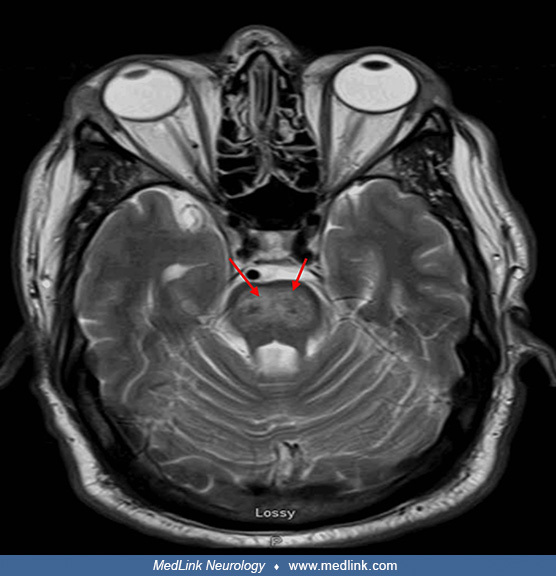
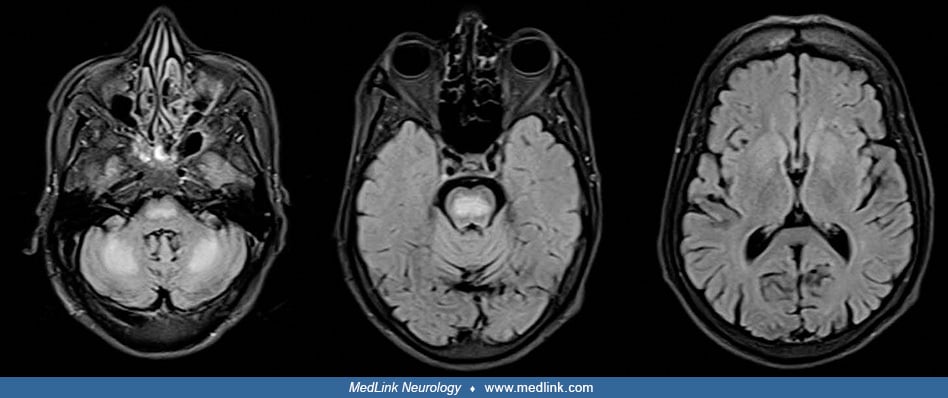
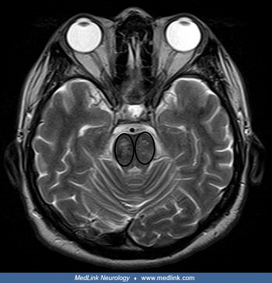
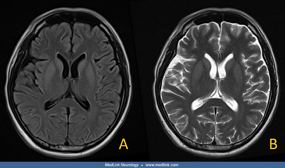
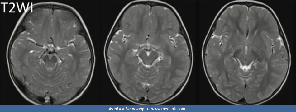
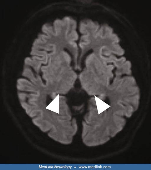
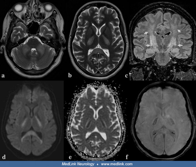
Axial T1-weighted MRI demonstrated symmetric low signal in the central pons (A, E). Axial T2-weighted MRI demonstrated a symmetric high signal in the central pons (B, F). T2 FLAIR images demonstrated a symmetric high signal in the central pons (C, G). Diffusion-weighted imaging (DWI) showed no diffusion restriction in the central pons (D, H). (Source: Jin X, Wang Y. Case report: osmotic demyelination syndrome after transcatheter aortic valve replacement: case report and review of current literature. Front Med (Lausanne) 2022;9:915981. Creative Commons Attribution license [CC BY], https://creativecommons.org/licenses/by/2.0.)