General Neurology
Lightning injuries: neurologic complications
May. 15, 2024
MedLink®, LLC
3525 Del Mar Heights Rd, Ste 304
San Diego, CA 92130-2122
Toll Free (U.S. + Canada): 800-452-2400
US Number: +1-619-640-4660
Support: service@medlink.com
Editor: editor@medlink.com
ISSN: 2831-9125
Toll Free (U.S. + Canada): 800-452-2400
US Number: +1-619-640-4660
Support: service@medlink.com
Editor: editor@medlink.com
ISSN: 2831-9125
Nearly 3,000 illustrations, including video clips of neurologic disorders.
Every article is reviewed by our esteemed Editorial Board for accuracy and currency.
Full spectrum of neurology in 1,200 comprehensive articles.
Listen to MedLink on the go with Audio versions of each article.
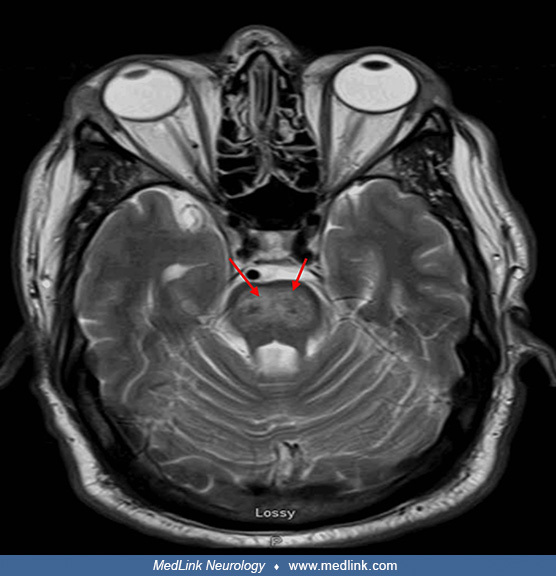
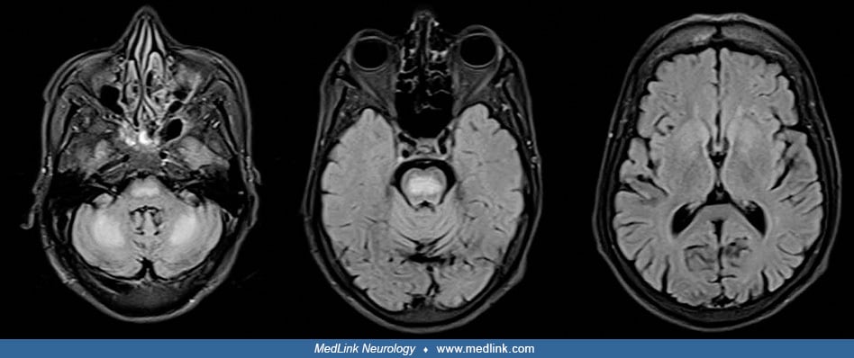
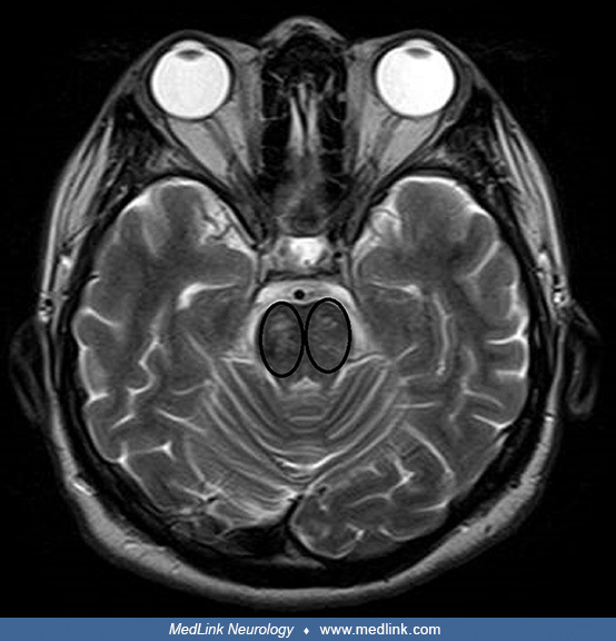
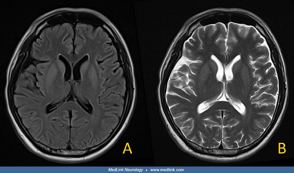
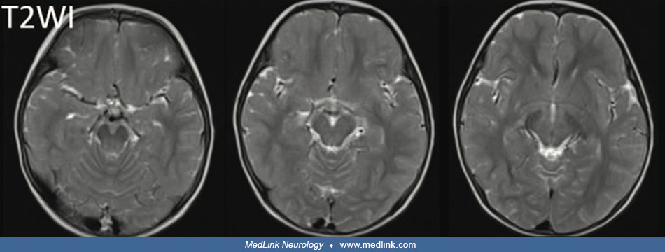
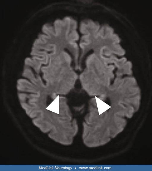
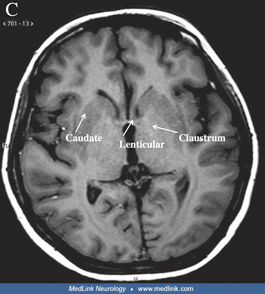
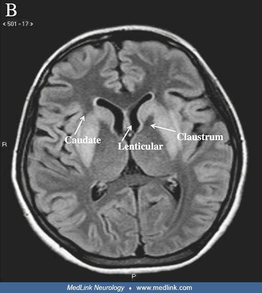
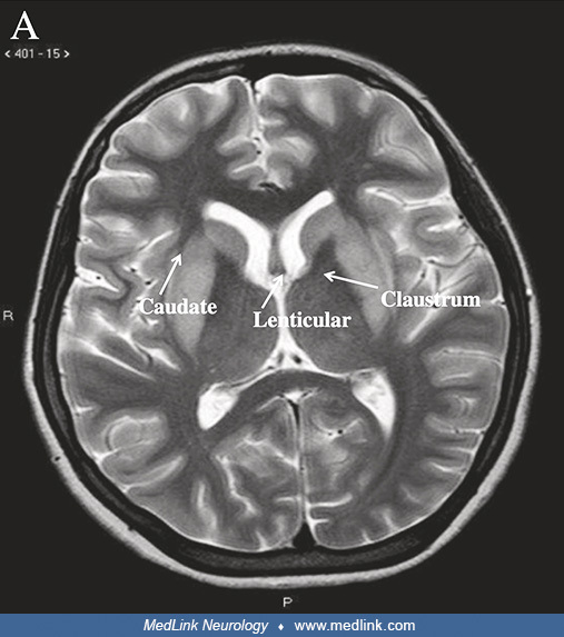
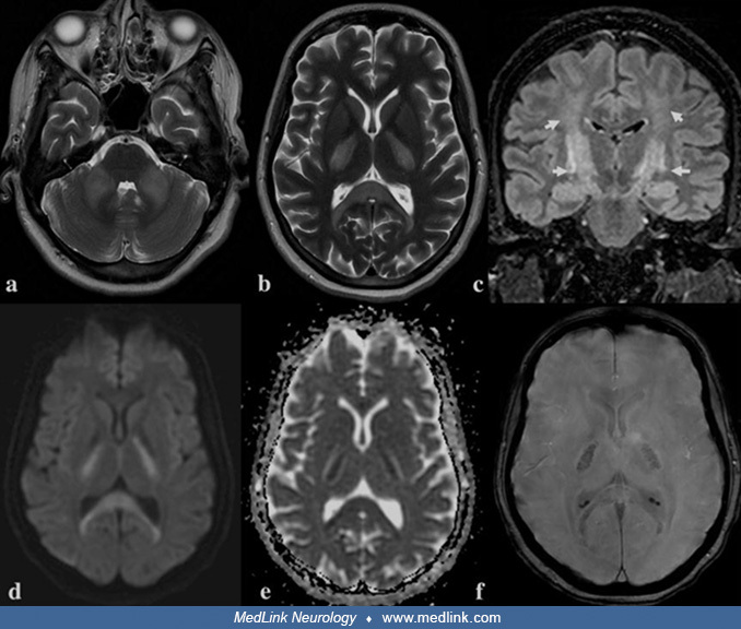
Axial T2 images (a, b) show abnormal hyperintense signal involving the middle cerebellar peduncles, splenium of the corpus callosum, and posterior limb of the internal capsule. The coronal fluid-attenuated inversion recovery (FLAIR) image (c) demonstrated selective involvement of the corticospinal tract (short arrows), giving a "wine-glass" appearance. The involved areas show restriction on diffusion-weighted images (d, e) with evidence of microhemorrhages on susceptibility-weighted images (f). The imaging features are characteristic of hypernatremic encephalopathy with osmotic myelinolysis and rhabdomyolysis. (Source: Choudhary G, Qureshi F, Arora A, Kothari N, Tiwari S, Bhatia P. Postpartum hypernatremia with extrapontine rhabdomyelinolysis: a case report. Qatar Med J 2022;2022[4]:45. Creative Commons Attribution license [CC BY], https://creativecommons.org/licenses/by/4.0.)